WB result of IMP3 Recombinant Rabbit mAb
Primary antibody: IMP3 Recombinant Rabbit mAb at 1/1000 dilution
Lane 1: HeLa whole cell lysate 20 µg
Lane 2: HEK-293 whole cell lysate 20 µg
Lane 3: BxPC-3 whole cell lysate 20 µg
Lane 4: Hep G2 whole cell lysate 20 µg
Lane 5: HT-29 whole cell lysate 20 µg
Secondary antibody: Goat Anti-Rabbit IgG (H+L), HRP conjugated at 1/10000 dilution
Predicted MW: 22 kDa
Observed MW: 69 kDa
IMP3 Recombinant Rabbit mAb (S-6196)
IMP3 Recombinant Rabbit mAb (S-6196)
Price:
Regular price
$100 USD
Regular price
Sale price
$100 USD
Unit price
per
For shipping services or bulk orders, you may request a quotation.
Secure checkout with

View full details
Product Details
Product Details
Product Specification
| Host | Rabbit |
| Antigen | IMP3 |
| Location | Nucleus |
| Accession | Q9NV31 |
| Clone Number | S-6196 |
| Antibody Type | Recombinant mAb |
| Isotype | IgG |
| Application | WB, IHC-P |
| Reactivity | Hu, Mk |
| Positive Sample | HeLa, HEK-293, BxPC-3, Hep G2, HT-29, COS-7 |
| Purification | Protein A |
| Concentration | 0.5 mg/ml |
| Conjugation | Unconjugated |
| Physical Appearance | Liquid |
| Storage Buffer | PBS, 40% Glycerol, 0.05% BSA, 0.02% sodium azide |
| Stability & Storage | 12 months from date of receipt / reconstitution, -20 °C as supplied |
Dilution
| application | dilution | species |
| WB | 1:1000 | Hu, Mk |
| IHC-P | 1:1000 | Hu |
Background
IMP3 (insulin-like growth factor 2 mRNA-binding protein 3, gene name IGF2BP3) is an oncofetal RNA-binding protein that is highly expressed during embryonic development and significantly downregulated after birth. It belongs to the IMP family, and its structure contains two N-terminal RNA recognition motifs (RRMs) and four C-terminal KH domains. As a reader of m6A modification, IMP3 regulates the stability, localization, and translational efficiency of transcripts by recognizing N6-methyladenosine-modified mRNAs, thereby exerting critical regulatory functions at the post-transcriptional level. IMP3 is aberrantly overexpressed in various malignant tumors, where it promotes cell proliferation, invasion, and metastasis by stabilizing oncogenic mRNAs such as MYC and CD44 or by activating IGF-2 translation, and it is closely associated with chemotherapy resistance and poor prognosis.
Picture
Picture
Western Blot
WB result of IMP3 Recombinant Rabbit mAb
Primary antibody: IMP3 Recombinant Rabbit mAb at 1/1000 dilution
Lane 1: COS-7 whole cell lysate 20 µg
Secondary antibody: Goat Anti-Rabbit IgG (H+L), HRP conjugated at 1/10000 dilution
Predicted MW: 22 kDa
Observed MW: 69 kDa
Immunohistochemistry
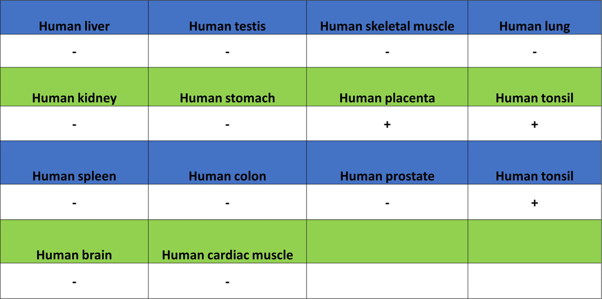
IHC shows positive staining in paraffin-embedded human tonsil. Anti-IMP3 antibody was used at 1/1000 dilution, followed by a HRP Polymer for Rabbit IgG (ready to use). Counterstained with hematoxylin. Heat mediated antigen retrieval with Tris/EDTA buffer pH9.0 was performed before commencing with IHC staining protocol.
IHC shows positive staining in paraffin-embedded human placenta. Anti-IMP3 antibody was used at 1/1000 dilution, followed by a HRP Polymer for Rabbit IgG (ready to use). Counterstained with hematoxylin. Heat mediated antigen retrieval with Tris/EDTA buffer pH9.0 was performed before commencing with IHC staining protocol.
Negative control: IHC shows negative staining in paraffin-embedded human kidney. Anti-IMP3 antibody was used at 1/1000 dilution, followed by a HRP Polymer for Rabbit IgG (ready to use). Counterstained with hematoxylin. Heat mediated antigen retrieval with Tris/EDTA buffer pH9.0 was performed before commencing with IHC staining protocol.
IHC shows positive staining in paraffin-embedded human gastric carcinoma. Anti-IMP3 antibody was used at 1/1000 dilution, followed by a HRP Polymer for Rabbit IgG (ready to use). Counterstained with hematoxylin. Heat mediated antigen retrieval with Tris/EDTA buffer pH9.0 was performed before commencing with IHC staining protocol.
IHC shows positive staining in paraffin-embedded human lung cancer. Anti-IMP3 antibody was used at 1/1000 dilution, followed by a HRP Polymer for Rabbit IgG (ready to use). Counterstained with hematoxylin. Heat mediated antigen retrieval with Tris/EDTA buffer pH9.0 was performed before commencing with IHC staining protocol.