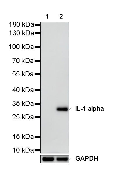
WB result of IL-1 alpha Recombinant Rabbit mAb
Primary antibody: IL-1 alpha Recombinant Rabbit mAb at 1/1000 dilution
Primary antibody incubation conditions: overnight at 4°C
Lane 1: untreated RAW 264.7 whole cell lysate 20 µg
Lane 2: RAW 264.7 treated with 5μg/mL LPS for 16 hours whole cell lysate 20 µg
Secondary antibody: Goat Anti-Rabbit IgG (H+L), HRP conjugated at 1/10000 dilution
Predicted MW: 31 kDa
Observed MW: 31 kDa
This blot was developed with high sensitivity substrate
IL-1 alpha Recombinant Rabbit mAb (S-6193)
IL-1 alpha Recombinant Rabbit mAb (S-6193)
Price:
Regular price
$100 USD
Regular price
Sale price
$100 USD
Unit price
per
For shipping services or bulk orders, you may request a quotation.
Secure checkout with

View full details
Product Details
Product Details
Product Specification
| Host | Rabbit |
| Antigen | IL-1 alpha |
| Synonyms | Interleukin-1 alpha; Il1a |
| Location | Cytoplasm, Nucleus, Secreted |
| Accession | P01582 |
| Clone Number | S-6193 |
| Antibody Type | Recombinant mAb |
| Isotype | IgG |
| Application | WB |
| Reactivity | Ms |
| Predicted Reactivity | / |
| Purification | Protein A |
| Concentration | 0.5 mg/ml |
| Conjugation | Unconjugated |
| Physical Appearance | Liquid |
| Storage Buffer | PBS, 40% Glycerol, 0.05% BSA, 0.02% sodium azide |
| Stability & Storage | 12 months from date of receipt / reconstitution, -20 °C as supplied |
Dilution
| application | dilution | species |
| WB | 1:1000 | Ms |
Background
Interleukin-1α (IL-1α) is a unique "dual-function" cytokine encoded by the IL1A gene. Unlike IL-1β, it is constitutively present in nearly all resting non-hematopoietic cells and serves a dual role as both a nuclear transcriptional regulator and an extracellular inflammatory signal. As a precursor protein (31 kDa), IL-1α is biologically active without requiring processing; its N-terminal pro-piece contains a nuclear localization sequence that allows it to enter the nucleus and participate in transcriptional regulation, while its C-terminal mature form (17 kDa) can be cleaved by the calcium-dependent protease calpain and secreted via a non-classical pathway. When cells undergo necrosis or tissue injury, IL-1α is passively released into the extracellular space, acting as an "alarmin" that rapidly initiates inflammatory responses, recruits neutrophils, and induces surrounding cells to produce IL-1β as well as other cytokines and chemokines. In addition, IL-1α plays a complex role in the tumor microenvironment: its membrane-bound form can enhance tumor immunogenicity, whereas the precursor form released by dying tumor cells promotes tumor invasion, angiogenesis, and myeloid cell-mediated immunosuppression. Its high nuclear expression is also associated with perineural invasion and poor prognosis in head and neck squamous cell carcinoma.
Picture
Picture
Western Blot