WB result of IFI16 Recombinant Rabbit mAb
Primary antibody: IFI16 Recombinant Rabbit mAb at 1/1000 dilution
Lane 1: HT-29 whole cell lysate 20 µg
Lane 2: Jurkat whole cell lysate 20 µg
Lane 3: Daudi whole cell lysate 20 µg
Lane 4: Raji whole cell lysate 20 µg
Negative control: HT-29 whole cell lysate
Secondary antibody: Goat Anti- rabbit IgG, (H+L), HRP conjugated at 1/10000 dilution
Predicted MW: 88 kDa
Observed MW: 100 kDa
IFI16 Recombinant Rabbit mAb (S-4089-44)
IFI16 Recombinant Rabbit mAb (S-4089-44)
Price:
Regular price
$100 USD
Regular price
Sale price
$100 USD
Unit price
per
For shipping services or bulk orders, you may request a quotation.
Secure checkout with

View full details
Product Details
Product Details
Product Specification
| Host | Rabbit |
| Antigen | IFI16 |
| Synonyms | Gamma-interferon-inducible protein 16; IFNGIP1 |
| Immunogen | Synthetic Peptide |
| Location | Cytoplasm, Nucleus |
| Accession | Q16666 |
| Clone Number | S-4089-44 |
| Antibody Type | Recombinant mAb |
| Isotype | IgG |
| Application | WB, ICC |
| Reactivity | Hu |
| Positive Sample | Jurkat, Daudi, Raji |
| Purification | Protein A |
| Concentration | 2 mg/ml |
| Physical Appearance | Liquid |
| Storage Buffer | PBS, 40% Glycerol, 0.05% BSA, 0.03% Proclin 300 |
| Stability & Storage | 12 months from date of receipt / reconstitution, -20 °C as supplied |
Dilution
| application | dilution | species |
| WB | 1:1000 | Hu |
| ICC | 1:2000 | Hu |
Background
Interferon gamma-inducible protein 16 (IFI16), also known as PYHIN1 or p204 in mice, is a crucial member of the PYHIN family of nuclear proteins that functions primarily as a innate immune sensor for foreign DNA within the nucleus of human cells. Characterized by an N-terminal pyrin domain and two C-terminal HIN-200 domains responsible for DNA binding, IFI16 detects double-stranded DNA from invading pathogens such as herpesviruses (e.g., KSHV, HSV-1) and retroviruses (e.g., HIV-1), triggering the assembly of inflammasomes or activating the STING-TBK1-IRF3 signaling axis to induce type I interferon and pro-inflammatory cytokine production. Beyond its role in pathogen recognition, IFI16 acts as a tumor suppressor by regulating cell cycle progression, promoting cellular senescence, and inducing apoptosis through interactions with key regulators like p53 and Rb, while its expression and activity are tightly modulated by post-translational modifications and viral countermeasures that often seek to degrade or sequester the protein to evade immune detection.
Picture
Picture
Western Blot
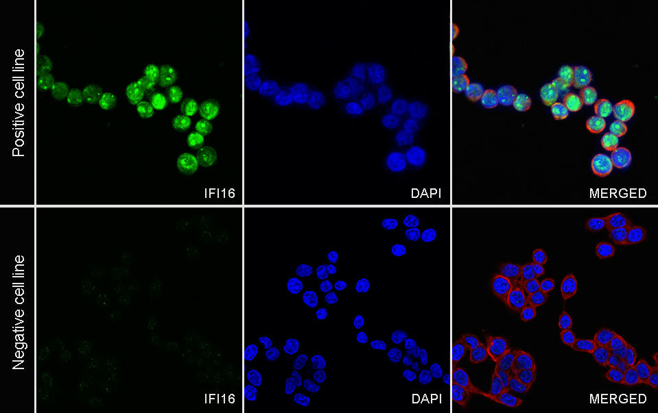
Immunocytochemistry
ICC shows positive staining in Jurkat cells (top panel) and negative staining in HT-29 cells (below panel). Anti-IFI16 antibody was used at 1/2000 dilution (Green) and incubated overnight at 4°C. Goat polyclonal Antibody to Rabbit IgG - H&L (Alexa Fluor® 488) was used as secondary antibody at 1/1000 dilution. The cells were fixed with 100% ice-cold methanol and permeabilized with 0.1% PBS-Triton X-100. Nuclei were counterstained with DAPI (Blue). Counterstain with tubulin (Red).