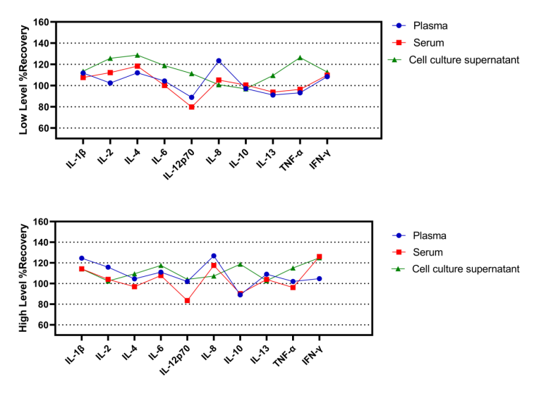
Linearity
After serial dilution of the high-concentration combined proteins of IL-1β,IL-2, IL-4, IL-6, IL-8,IL-10, IL-12p70,IL-13,TNF-α,and IFN-γ using plasma, serum, and cell supernatant matrices respectively, the linear slopes between the actual detected values and the theoretical values are as shown in the table.
Human Th1/Th2/Inflammation 10-Plex Assay (Luminex)
Human Th1/Th2/Inflammation 10-Plex Assay (Luminex)
Price:
Regular price
$4,000 USD
Regular price
Sale price
$4,000 USD
Unit price
per
For shipping services or bulk orders, you may request a quotation.
Secure checkout with

View full details
Product Details
Product Details
Product Specification
| Antigen | IL-1β、IL-2、IL-4、IL-6、IL-8、IL-10、IL-12p70、IL-13、TNF-α、IFN-γ |
| Antibody Type | Recombinant mAb |
| Reactivity | Human |
| Stability & Storage | 12 months from date of receipt / reconstitution, 2 to 8℃ as supplied. |
Kit
| Precision | Intra-assay coefficient of variation: <10%; Inter-assay coefficient of variation: <15% |
| Sample type | Cell culture supernatant, Serum, Plasma |
| Assay type | Sandwich (Qualitative) |
| Sensitivity | <1.0pg/ml |
| Range | 0.2-10000 pg/mL |
| Recovery | 70%-130% |
| Assay time | 70 minutes |
| Species reactivity | Human |
| Plate | Non-detachable plate |
Background
xMAP Technology uses colored beads to perform biological assays similar to ELISA or nucleic acid hybridization assays. By color-coding microscopic beads into many spectrally distinct sets, each bead set can be coated with a nucleic acid or protein capture molecule specific to a particular biological target, allowing the simultaneous capture of multiple analytes from a single sample. Because of the microscopic size and low density of these beads, assay reactions exhibit virtually solution-phase kinetics. However, once an assay is complete the solid phase characteristics allow each bead to be analyzed discretely. By incorporating magnetic properties into xMAP microspheres, assay washing is simplified while maintaining desirable solution-phase properties.
Picture
Picture
Luminex
Standard Curve
Example of IL-1β,IL-2, IL-4, IL-6, IL-8,IL-10, IL-12p70,IL-13,TNF-α,and IFN-γ standard curve in Assay Diluent.
Quantifiction of Human PBMC
PBMC Unsimulated Supernatant: The cells of PBMCs were cultured for24 hours, the cell culture supernatant was collected and analyzed quantitatively using S0X1004.
PBMC simulated Supernatant: The cells of PBMCs were stimulated with 10 μg/mL PMA. After 24 hours, the cell culture supernatant was collected and analyzed quantitatively using S0X1004.
Intra-assay precision
Ten replicates of each of three different levels of IL-1β,IL-2, IL-4, IL-6, IL-8,IL-10, IL-12p70,IL-13,TNF-α,and IFN-γ were tested.
Inter-assay precision
Ten repeated tests were conducted on three different levels of IL-1β,IL-2, IL-4, IL-6, IL-8,IL-10, IL-12p70,IL-13,TNF-α,and IFN-γ using different batches of reagents.
Specificity
The antibodies used in the kit have been screened for their specific reactivity with particular cytokines. After analyzing samples containing only a single recombinant cytokine protein, it was found that when using this detection method, there was no cross-reaction or background detection of cytokines in other capture magnetic bead groups.
Theoretical limit of detection
Repeat the detection of the blank sample 20 times using the same batch of reagents, and calculate the theoretical limit of detection.
Protocol Diagram
Recovery
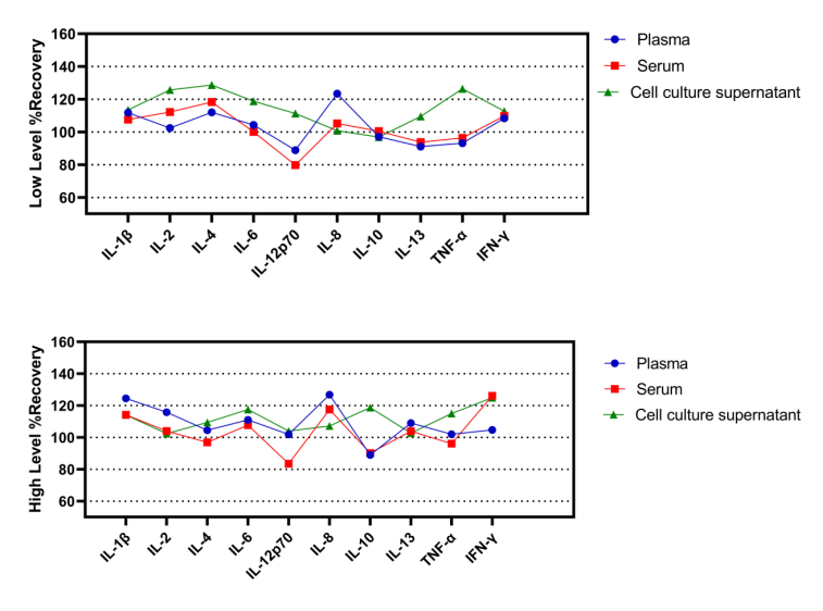
Within the detection range, the combined cytokine proteins were added to serum,plasma,cell supernatant at high concentrations.The matrices used in these experiments were diluted 2-fold with the assay buffer before the addition of the cytokine proteins. Theresults were compared with those of samples that had the same concentration of cytokines added in the assay buffer.