Human Total IgG Kit (High Sensitivity) (HICA)
Human Total IgG Kit (High Sensitivity) (HICA)

Product Details
Product Details
Product Specification
| Stability & Storage | Store at 2–8°C, protected from light, for 6 months. After reconstitution, the calibrator may be aliquoted and stored at −20°C for up to 3 months; avoid repeated freeze-thaw cycles. |
Background
Human immunoglobulinG (Human Immunoglobulin G, IgG) is an antibody secreted and released by plasma cells (B cells). It is the most common type of antibody in the bloodstream and the most abundant immunoglobulin in human serum, accounting for approximately 75% of total serum immunoglobulins. The molecular weight of IgG is about 150 kDa, and it typically exists as a tetrameric structure composed of two identical heavy chains and two identical light chains linked by interchain disulfide bonds. IgG serves as a major effector molecule in humoral immune responses. Based on differences in the amino acid composition and structure of the hinge region, IgG can be divided into four subclasses: IgG1 (approximately 66%), IgG2 (approximately 23%), IgG3 (approximately 7%), and IgG4 (approximately 4%). IgG is distributed in blood and tissue fluids, where it exerts various immune functions, including neutralizing pathogens, activating complement, opsonizing phagocytosis, and mediating antibody-dependent cell-mediated cytotoxicity. It plays a central role in anti-infection immunity and immunological memory and can cross the placenta to provide passive immune protection to newborns. Clinically, IgG is an important indicator for assessing humoral immunity, vaccine efficacy, and the mechanisms of autoimmune diseases, and it is widely used in disease mechanism research, drug efficacy evaluation, and biomarker development. This kit…For the quantitative in vitro detection of total [substance] in cell culture supernatants, serum, plasma, and other samples.IgG concentration provides a sensitive, specific, and efficient tool suitable for basic medical research and drug screening applications.
Principle of the test:
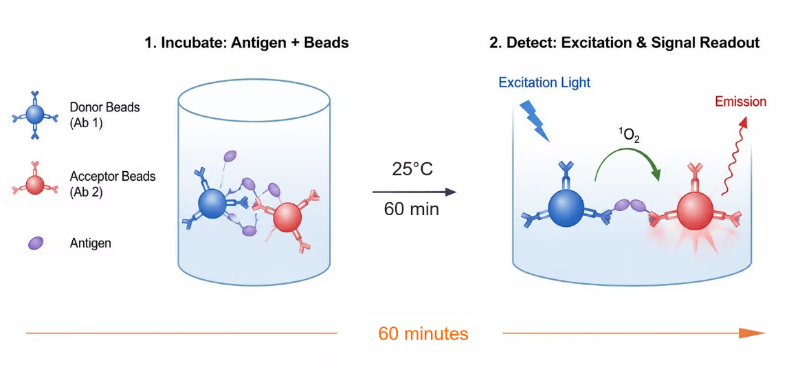
This kit employs a homogeneous chemiluminescence assay based on the sandwich ELISA method.Homogeneous Immuno Chemiluminescence Assay, HICA) to detect cytokine concentrations, with simple operation,No washing required.
The detection system includes two types of microspheres:DonorMicrosphere conjugation targeting the target proteinAntibody 1,ReceptorMicrosphere conjugationAntibody to the target protein 2At the time of the reaction, the target protein binds to the antibody and the microspheres, forming an immune complex that brings the two types of microspheres into close proximity. When the distance between the donor microsphere and the acceptor microsphere is less than 200 nm, the singlet oxygen generated upon light excitation can be transferred to the acceptor microsphere, triggering chemiluminescence; conversely, if the target protein is absent and the microspheres are too far apart, no signal is produced.
Quantitative analysis of the target protein can be achieved by measuring the intensity of the chemiluminescent signal. This method is characterized by its simplicity, rapid reaction kinetics, and high sensitivity.
Components
Name |
Specification |
Component Specification |
Test ReagentR1 |
100 Tests |
6μl /per tube ×1 |
500 Tests |
30μl /per tube ×1 |
|
Test ReagentR2 |
100 Tests |
6μl /per tube ×1 |
500 Tests |
30μl /per tube ×1 |
|
Calibrator |
100 Tests |
100 ngLyophilized powder ×1 |
500 Tests |
100 ngLyophilized powder ×5 |
|
Reagent Diluent |
100 Tests |
2.2 mL/per bottle ×1 |
500 Tests |
11 mL/per bottle ×1 |
|
Calibrator Diluent |
100 Tests |
1 mL/per tube ×1 |
500 Tests |
1 mL/per tube ×5 |
Note: It is recommended to use microplates for the assay (384shallow wellsor384,96well plates, white or gray)
Protocol
Sample requirements
1.To remove impurities from the sample, the sample must be centrifuged prior to analysis.2000 ×g,10minutes).
2.If the measured concentration of a sample exceeds the highest value on the standard curve, it should be appropriately diluted (using negative serum is recommended) and retested to ensure that the result falls within the valid measurement range.
Testing process
2.1Preparation of calibration standard gradient samplesAdopt 100muLReconstitute with deionized water.100The lyophilized calibrator is designated as C.12, then dilute the reconstituted lyophilized calibrator C with the calibrator diluent.12Perform gradient dilution, calibrator C.1This is the calibrator diluent; the recommended dilution protocol is shown in the table below:
Gradient |
Concentration (pg/mL) |
Higher-level schoolQualified product(microliters) |
Diluent (μL) |
C12 |
1,000,000 |
- |
- |
C11 |
300,000 |
30μL C12 |
70 |
C10 |
100000 |
30μL C11 |
60 |
C9 |
30000 |
30μL C10 |
70 |
C8 |
10000 |
30μL C9 |
60 |
C7 |
3000 |
30μL C8 |
70 |
C6 |
1000 |
30μL C7 |
60 |
C5 |
300 |
30μL C6 |
70 |
C4 |
100 |
30μL C5 |
60 |
C3 |
30 |
30μL C4 |
70 |
C2 |
10 |
30μL C3 |
60 |
C1 |
0 |
- |
- |
2.2Testing procedure:
|
Testing procedure 25℃One-step methodTesting | |
Sample addition |
1.Take2µL of sampleAdd to384microplate wells; 2.UltrasonicateR1andR2for5 secondsAfter vortexing or mixing, mix equal volumes and homogenize, then dilute with the reagent diluent.200fold and mix to prepare the working reagent solution; 3.Add to the sampleadd18µL of the premixedworking reagent solution |
incubate |
on a microplate shaker800 r/min25°Cincubate60min |
read the results |
instrument reading,protect from light/green light |
Note:the dilutedR1andR2should be used withinhalfan hour after mixing, and must beprotected from light/green light;
the reaction system can be adjusted proportionally according to experimental needs.
III. Performance Testing
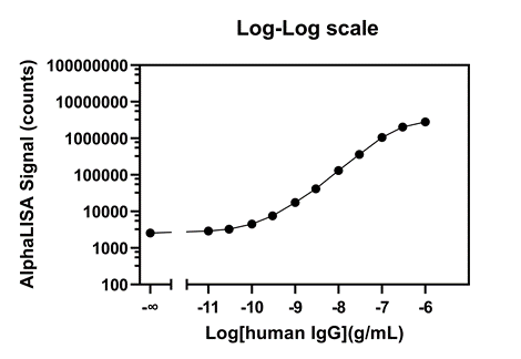
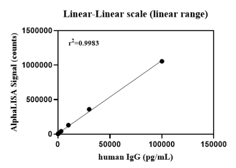
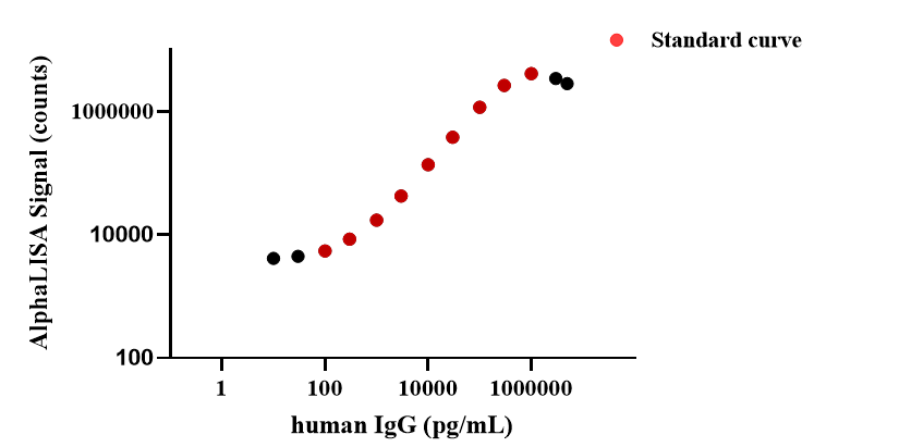
3.1Example of a complete standard curve:
3.2Performance parameter verification:
LOB:repeat the detection20times for the calibration standardC1, calculate the mean signal andSD, use the standard curve to calculate the concentration value corresponding tomean signal+2×SD, which is the limit of detection LOB.
matrix |
blank limit(pg/mL) |
buffer |
7.0 |
dynamic range (Dynamic range):0 –1000000 pg/mL.
Linear range (Linear Range):100– 100000 pg/mL.
Precision (Precision):
Intra-precision-Precision): In the same experimentOn the orifice plate, using known low,China,Each high-concentration sample was tested in triplicate.1Zero times, to evaluate the intra-assay precision of this assay.schoolBoth the reference product and the sampleTest using different processes.The coefficient of variation (%CV) for the test results of samples at each concentration was less than 10%, indicating that the method exhibits good reproducibility within the same batch..
Matrix |
Precision |
||
lowConcentration |
Medium concentration |
High concentration |
|
Buffer solution |
4% |
3% |
1% |
Inter-plate precision (Inter-Precision): In 5In the next independent experiment, known low levels were tested separately,China,High-concentration samples are being tested. Settings5repeated times. The test results of samples at each concentration all showed a low coefficient of variation (CV < 10%), indicating that the method has good reproducibility between different batches.
Matrix |
Intermediate precision |
||
Low concentration |
Medium concentration |
High concentration |
|
Buffer |
8% |
5% |
4% |
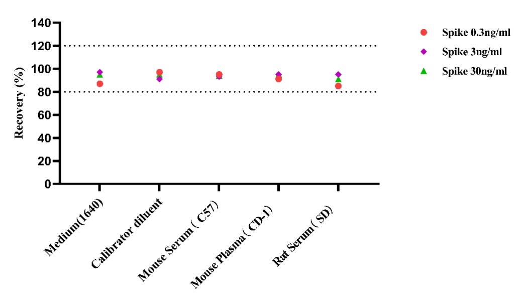
Accuracy (Recovery):
Standard solutions of IgG at low, medium, and high concentrations were added to different matrices, and their average recovery rates were tested twice.IgGMatrices
Recovery rate |
Low concentration |
||
Medium concentration |
High concentration |
Calibrator diluent |
|
97% |
91% |
95% |
Culture supernatant |
87% |
97% |
95% |
200X |
Mouse serum95 |
%93% |
94% |
200X |
Mouse plasma91% |
95% |
95% |
95% |
200XRat serum |
85% |
95% |
91% |
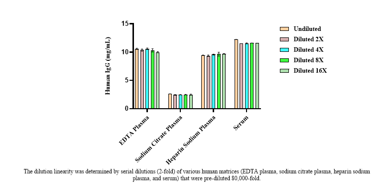
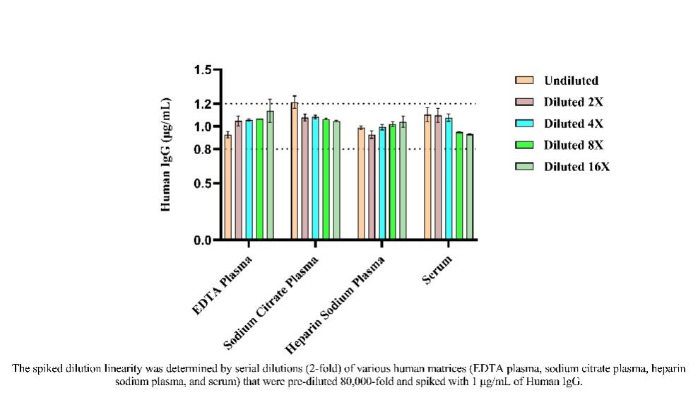
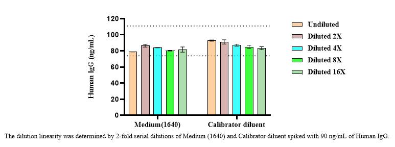
Dilution linearity (Dilution linearity):
The samples were diluted by factors of 8The serum and plasma were diluted in a gradient after being multiplied by a factor, and corrections were applied; additionally, the supernatant of the culture medium, calibrator diluent, and dilutions were used.Serum and plasma after 80,000-fold dilutionAdd to the middleIgGThe standard was serially diluted and used for calibration to evaluate its dilution linearity.
Hook effect (HOOK):
takeIgGStandard substanceGradient dilution and testing confirmed that samples within the measurement range yield reliable quantitative results, and no signal suppression was observed.
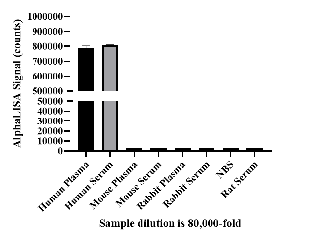
Specificity (Specificity:):
Dilute sera and plasmas from different species separately.After 80,000-fold dilution, the results showed that this kit exhibited no cross-reactivity in the following species.
Guidelines
Reagents R1 and R2 should be used protected from light; sample addition and incubation are recommended to be performed under green light or dim light (100 LUX).
Each assay requires recalibration, with each concentration level of the calibrator tested in at least duplicate wells, using five-parameter curve fitting for calculation.
During incubation, temperature and time must be strictly controlled; it is recommended to cover the microplate with a film, and an ELISA reader with ALPHA functionality should be used.
Components from different lot numbers of the kit must not be mixed.