UniOne® TR-FRET Human STAT6/CRBN PROTAC Binding Kit
UniOne® TR-FRET Human STAT6/CRBN PROTAC Binding Kit

Product Details
Product Details
Product Specification
| Host | Human |
| Stability & Storage | -80℃ |
Background
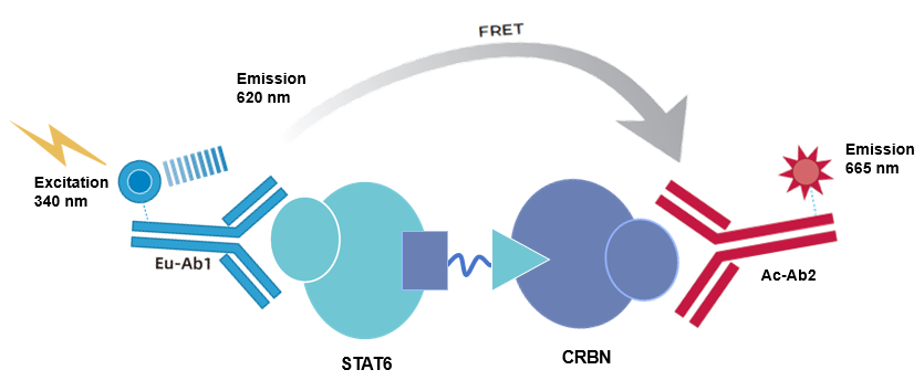
The kit employs homogeneous time-resolved fluorescence (TR-FRET) technology to measure the interaction between molecular glue AK-1690 and test compounds mediating STAT6 and CRBN. This method enables high-throughput screening of small molecules capable of mediating the interaction between STAT6 and CRBN in a simple and rapid manner.
As shown, the interaction between STAT6 and CRBN is detected using Eu-labeled anti-Tag1 antibody (TR-FRET donor) and Ac-labeled anti-Tag2 antibody (TR-FRET acceptor). Since molecular glue AK-1690 mediates the interaction between STAT6 and CRBN, the proximity of the donor and acceptor antibodies allows fluorescence resonance energy transfer (FRET) from the donor to the acceptor upon excitation, resulting in a specific emission signal at 665 nm. This signal is proportional to the extent of interaction mediated by AK-1690 between STAT6 and CRBN. The homogeneous assay is simple to perform and requires no washing steps.
Components
Component |
Concentration |
100T |
500T |
2500T |
10000T |
Storage Temperature |
Tag1-STAT6 protein |
50× |
10μL |
40μL |
200μL |
800μL |
-80℃ |
Tag2- CRBN protein |
50× |
10μL |
40μL |
200μL |
800μL |
-80℃ |
AK-1690 (150μM) |
100× |
5μL |
10μL |
50μL |
200μL |
-80℃极p> |
Anti-Tag1 Eu antibody |
50× |
10μL |
50μL |
250μL |
1000μL |
-80℃ |
Anti-Tag2 Ac antibody |
12.5× |
40μL |
200μL |
1000μL |
4000μL |
-80℃ |
Detection buffer |
10× |
400μL |
2mL |
10mL |
40mL |
-80℃ |
Protocol
1. Reagent Preparation
1.1 Thaw all reagents at room temperature before use (equilibrate at room temperature for at least 30 min). The reaction volume for the 384-well shallow plate is 20μL (reagent volumes for the reaction system are shown in the table). Calculate the required volume for the experiment before preparation and prepare as needed; the following preparation is for reference only, using 500 assays as an example.
Table 1. Reagent Preparation
Reagent Name |
Preparation |
Volume per Assay Well (μL) |
Detection buffer 1 |
Take2mL 10 × Detection buffer1and add18mL deionized water to diluteto1×. Mixwell and setaside. |
- |
AK-1690 |
According to the reaction system, dilute the compound with1×Detection buffer to the desired testing concentration. Ensure consistentDMSO concentration across all assay wells. |
2 |
Tag1-STAT6 protein |
Take40μL Tag1-STAT6 protein stock solution and dilute to 2mL with 1× Detection buffer.Mix well and set aside. |
4 |
Tag2- CRBN protein |
Take40μL Tag2-CRBN protein stock solution and dilute to 2mL with 1×Detection buffer.Mix well and set aside. |
4 |
Antibody Mix |
Take50μL Anti-Tag1 Eu antibody stock solution and add 1×Detection buffer 2.45mL. Mix well;take200μL Anti-Tag2 Ac antibody stock solution and add 1× Detection buffer 2.3mL. Mix well;mix the two solutions at a1:1 ratio to prepare the Antibody Mix. |
10 |
1.2 Gradient Dilution of Test Samples
Using AK-1690 as an example, the diluent is 1× Detection buffer. To minimize the impact of matrix effects, it is recommended to dilute with a solution matching the matrix of the test sample; adjust the test sample concentrations according to actual conditions.
Table2. AK-1690 Gradient Dilution (Adjust according to actual conditions)
Final Concentration of AK-1690 (nM) |
Preparation Concentration of AK-1690 (nM) |
Preparation Method |
|
① |
150 |
1500 |
1μL 150μM AK-1690 +99μL 1× Detection buffer |
② |
100 |
1000 |
30μL ① +15μL 1× Detection buffer |
③ |
66.67 |
666.67 |
30μL ② +15μL 1× Detection buffer |
④ |
44.44 |
444.44 |
30μL ③ +15μL 1× Detection buffer |
⑤ |
29.63 |
296.30 |
30μL ④ +15μL 1× Detection buffer |
⑥ |
19.75 |
197.53 |
30μL ⑤ +15μL 1× Detection buffer |
⑦ |
13.17 |
131.69 |
30μL ⑥ +15μL 1× Detection buffer |
⑧ |
8.78 |
87.79 |
30μL ⑦+15μL 1× Detection buffer |
⑨ |
5.85 |
58.53 |
30μL ⑧+15μL 1× Detection buffer |
Blank |
0 |
0 |
30μL 1× Detection buffer |
2. Sample Addition and Controls
2.1 Test Samples: Add 2μL of gradient-diluted test sample, 4μL of Tag1-STAT6 protein working solution, 4μL of Tag2-CRBN protein working solution, and 10μL of mixed Antibody Mix (Tag2-CRBN protein can be mixed with Antibody Mix at a proportional ratio before addition) sequentially into the 384-well shallow plate.
2.2 Positive Control Standard Curve: 2μL of gradient-diluted AK-1690, 4μL of Tag1-STAT6 protein working solution, 4μL of Tag2-CRBN protein working solution, and 10μL of Antibody Mix.
Positive Control Standard Curve |
Test Samples |
Blank Control Wells |
NC |
|
Step 1 |
2μL Gradient-dilutedAK-1690 |
2μL Gradient-diluted Test Samples |
2μL 1× Detection buffer |
10μL 1× Detection buffer Add 10μL Antibody Mix |
4μL Tag1- STAT6 protein | ||||
Step 2 |
4μL Tag2-CRBN protein |
|||
10μL Antibody Mix | ||||
Seal the plate wells with a sealing film. Incubate at room temperature for 2 hours; | ||||
2.3 Blank Control Wells: Replace test samples with 2μL of 1× Detection buffer;
2.4 NC: 10μL of 1× Detection buffer plus 10μL of Antibody Mix.
After adding all samples, centrifuge, seal with a sealing film, and incubate at room temperature for 2 hours.
3. Detection
Detect using a microplate reader compatible with TR-FRET. Excitation wavelength is 320/340nm, and emission wavelengths are 620nm and 665nm.
[Result Calculation]
1) Calculate the signal value (Ratio): Divide the 665nm fluorescence signal by the 620nm fluorescence signal, then multiply by 10000.
Ratio = (665/620) ×10000
2) CalculateNet signal based on the signal value:
Net signal = (Std-NC)/NC×100
3) Calculate CV(%):
CV(%)= Standard Deviation/Mean Ratio × 100%
[Data Example]
The following data cannot replace data obtained from actual experiments and is provided for illustration only. Results may vary depending on the plate reader.
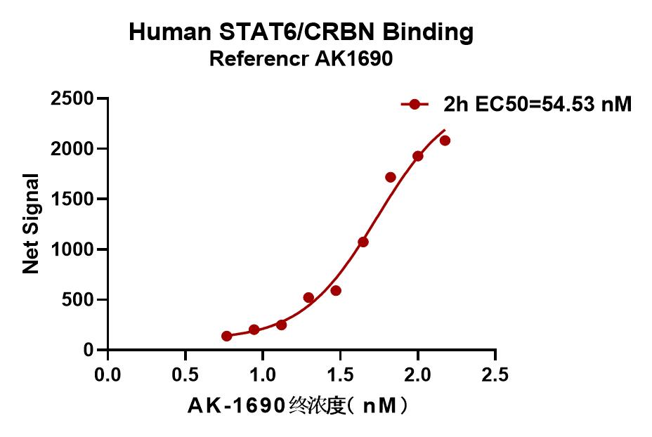
Note: Recommended microplate (384-well plate, white, shallow well)