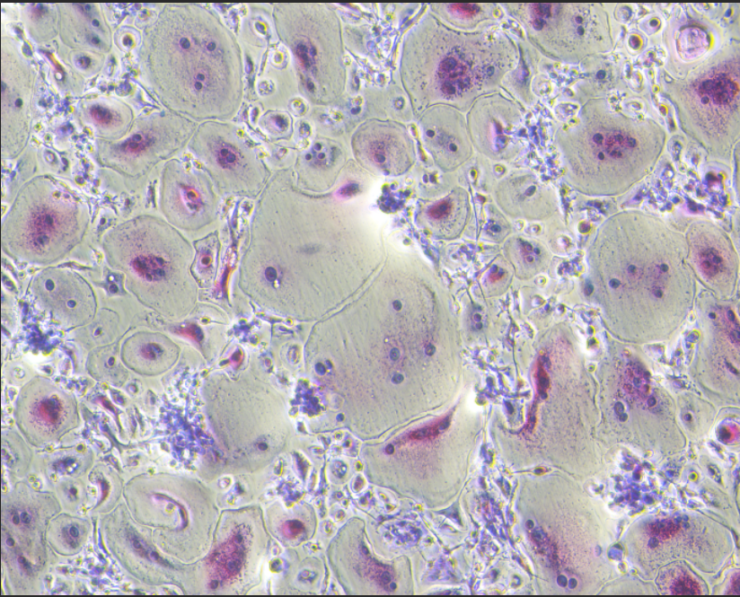
After 6 days of RANKL induction, the classic multinucleated osteoclast structure can be observed under a microscope.
Human Monocyte-Induced Osteoclast Differentiation Cytokine Kit
Human Monocyte-Induced Osteoclast Differentiation Cytokine Kit
Price:
Regular price
$245 USD
Regular price
Sale price
$245 USD
Unit price
per
For shipping services or bulk orders, you may request a quotation.
Secure checkout with

View full details
Product Details
Product Details
Product Specification
| Synonyms | Human Monocyte-Induced Osteoclast Differentiation Cytokine Kit |
| Endotoxin | <0.1EU/μg |
| Physical Appearance | Lyophilized powder |
| Reconstitution | Reconstitute at 0.1-1 mg/ml according to the size in ultrapure water after rapid centrifugation. |
| Stability & Storage | ·12 months from date of receipt, lyophilized powder stored at -20 to -80℃. |
Components
Catalog Number |
Protein |
Reference dosage |
S Size(100ml System) |
M Size(1000ml System) |
UA040042 |
M-CSF Protein, Human |
50ng/mL |
5μg |
50μg |
UA040329 |
RANK L/TNFSF11(178aa) Protein, Human |
100ng/mL |
10μg |
100μg |
Protocol
Preparation of Human Peripheral Blood Monocyte-Derived Dendritic Cells:
1. Isolation of CD14⁺ Monocytes
1.1 Thaw cryopreserved PBMCs and centrifuge at 300 × g for 5 min at room temperature. Aspirate the supernatant and resuspend the cells pellet in complete α-MEM medium [ containing 10% fetal bovine serum (FBS) and 1% penicillin-streptomycin]. Perform cell counting and viability assessment (cell viability should be > 95%). Incubate the cells with CD14 Beads (human, Cat. No. S0K0004) for positive selection according to the manufacturer's protocol. Isolate and collect the CD14⁺ cell fraction using a magnetic separator. The purity of the isolated CD14⁺ population should be verified by flow cytometry using a small aliquot. A purity greater than 95% is recommended to ensure the quality of subsequent steps.
2. Osteoclast Differentiation Induction
2.1 Use complete α-MEM medium (containing 10% FBS and 1% penicillin-streptomycin) to adjust the density of sorted CD14+ cells to 2 × 10⁵ cells/mL. Seed the cells into a 24-well plate at a volume of 1 mL per well. Immediately add the recombinant human cytokine M-CSF (50 ng/mL), and place the 24-well plate in a 37°C, 5% CO₂ incubator for cultivation.
2.2 Replace with fresh complete medium every 2-3 days. During the medium replacement, gently aspirate the old medium and add an equal volume of fresh complete medium containing the same concentration of M-CSF. After 7 days, supplement the medium with RANKL (100 ng/mL) to initiate the osteoclast differentiation process. Thereafter, maintain the synergistic induction of both M-CSF and RANKL factors. During this period, replace the complete medium every 2-3 days with fresh complete medium containing the same concentrations of M-CSF and RANKL. By days 12-14, large multinucleated giant cells should be visible under the microscope, which are TRAP-positive and possess bone resorption function, indicating successful induction of mature osteoclasts.
3. Cell Morphological Observation and Identification
3.1 Morphological Observation: Starting from the initiation of induction, regularly observe the morphological changes of the cells under an inverted microscope. Typically, characteristic multinucleated osteoclast-like structures can be observed 5 days after RANKL induction.
3.2 TRAP Staining Validation: After 5-7 days of synergistic induction with M-CSF and RANKL, perform Tartrate-Resistant Acid Phosphatase (TRAP) staining to specifically identify osteoclasts. Strictly follow the staining procedure steps provided in the TRAP staining kit instruction manual. Once staining is completed, observe the cells under a light microscope. Mature osteoclasts are defined as TRAP-positive cells (exhibiting red or purplish-red cytoplasm) that contain 3 or more nuclei.
Guidelines
Store in separate containers to minimize freeze-thaw cycles.
Picture
Picture
Bioactivity
After 6 days of RANKL induction, tartrate-resistant acid phosphatase (TRAP) staining was performed to specifically identify osteoclasts. After staining, the mature osteoclasts were observed under an optical microscope, and their cytoplasm appeared red or reddish purple.