Standard curve
Example of Human Hemoglobin standard curve in Assay Diluent DC4K7.
Human & Monkey Hemoglobin culture media OneStep ELISA Kit
Human & Monkey Hemoglobin culture media OneStep ELISA Kit
Price:
Regular price
$500 USD
Regular price
Sale price
$500 USD
Unit price
per
For shipping services or bulk orders, you may request a quotation.
Secure checkout with

View full details
Product Details
Product Details
Product Specification
| Antigen | Hemoglobin |
| Immunogen | Recombinant Protein |
| Antibody Type | Recombinant mAb |
| Reactivity | Hu, Mk |
| Purification | Protein A |
| Stability & Storage | 12 months from date of receipt / reconstitution, 2 to 8°C as supplied. |
Kit
| Precision | Intra-assay: 1.5% Inter-assay: 3.3% |
| Sample type | Cell culture supernatant |
| Assay type | Sandwich (quantitative) |
| Sensitivity | 2.627 ng/mL |
| Range | 5.27 ng/mL – 60 ng/mL |
| Recovery | Cell culture supernatant: 105% |
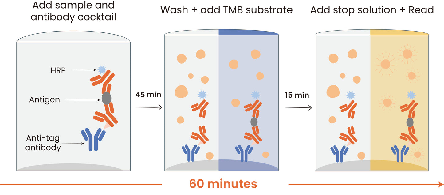
| Assay time | 60 minutes |
| Species reactivity | Hu; Mk |
Background
Human hemoglobin is a complex metalloprotein found in red blood cells that is essential for transporting oxygen from the lungs to body tissues and facilitating the return transport of carbon dioxide. Structurally, it is a tetramer consisting of two alpha and two beta polypeptide chains in adults (HbA), each bound to a heme group containing a ferrous iron atom that can reversibly bind one oxygen molecule, allowing a single hemoglobin molecule to carry up to four oxygen molecules. Functionally, hemoglobin exhibits cooperative binding, where the binding of one oxygen molecule increases the affinity for the next, resulting in a sigmoidal oxygen dissociation curve modulated by factors such as pH (the Bohr effect), carbon dioxide, and 2,3-bisphosphoglycerate (2,3-BPG) to adapt oxygen release to tissue metabolic needs. Mutations in the globin genes can lead to disorders like sickle cell disease and thalassemias, making the understanding of hemoglobin’s structure and function fundamental to both physiology and clinical medicine.
Picture
Picture
ELISA
To assess the linearity of the assay, the sample was spiked with high concentrations of Human hemoglobin in various matrices and diluted with the appropriate Calibrator Diluent to produce samples with values within the dynamic range of the assay.
Protocol Diagram