Standard curve
Example of IL-7 standard curve in Assay Diluent .
Human IL-7 Single Plex Assay (适用于Luminex)
Human IL-7 Single Plex Assay (适用于Luminex)
Price:
Regular price
$585 USD
Regular price
Sale price
$585 USD
Unit price
per
For shipping services or bulk orders, you may request a quotation.
Secure checkout with

View full details
Product Details
Product Details
Product Specification
| Antigen | IL-7 |
| Reactivity | Human |
| Stability & Storage | 12 months from date of receipt / reconstitution, 2 to 8°C as supplied. |
Kit
| Precision | Intra-assay: <10%; Inter-assay: <15% |
| Sample type | Serum; Plasma; Cell culture supernatant |
| Assay type | Sandwich (quantitative) |
| Sensitivity | <0.5 pg/mL |
| Range | 0.3pg/mL-4500pg/mL |
| Recovery | Plasma: 80%-120% Serum: 80%-120% Cell culture supernatant: 70%-130% |
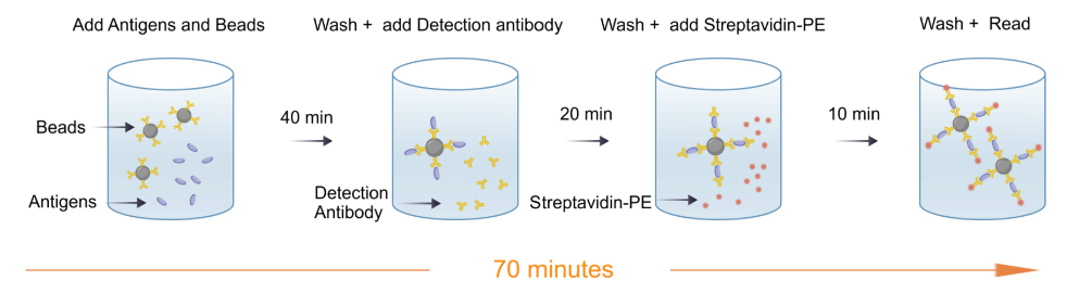
| Assay time | 70 minutes |
| Species reactivity | Human |
Background
IL7, also known as interleukin 7, is a hematopoietic growth factor that belongs to the IL-7/IL-9 family. It is secreted by stromal cells in the bone marrow and thymus. It is also produced by keratinocytes, dendritic cells, hepatocytes, neurons, and epithelial cells, but is not produced by lymphocytes. Human IL7 cDNA encodes 177 amino acids (aa) that include a 25 aa signal peptide. Human IL-7 shares approximately 60-63% aa sequence identity with mouse, rat, canine and feline IL-7, and 72-76% with equine, bovine, ovine, and porcine IL-7. IL7 stimulates the proliferation of lymphoid progenitors. It is important for proliferation during certain stages of B-cell maturation. Like other common gamma-chain cytokines like IL-2 and IL-15, IL-7 and its receptor, IL-7R, have been used in a variety of immunotherapy applications, often in fluid tumors and in some instances of solid tumor models.
Picture
Picture
Luminex
Quantifiction of human PBMC
Stimulated sample: Human peripheral blood mononuclear cells (PBMCs) were stimulated with CD3、PMA、PHA、LPS、Poly(I:C), and the average concentration of IL-7 in the supernatant was measured to be 16.23 pg/mL.
Unstimulated sample: Human PBMCs were cultured for 24h, and the average concentration of IL-7 in the supernatant was measured to be 13.47 pg/mL.
Recovery
Within the detection range, the combined cytokine proteins were added to serum,plasma,cell supernatant at high、low concentrations.The matrices used in these experiments were diluted 2-fold with the assay buffer before the addition of the cytokine proteins. The results were compared with those of samples that had the same concentration of cytokines added in the assay buffer.
Protocol Diagram