Standard curve
Example of IL-3 standard curve in Assay Diluent .
Human IL-3 Single Plex Kit (Flow Cytometry Bead Assay)
Human IL-3 Single Plex Kit (Flow Cytometry Bead Assay)
Price:
Regular price
$500 USD
Regular price
Sale price
$500 USD
Unit price
per
For shipping services or bulk orders, you may request a quotation.
Secure checkout with

View full details
Product Details
Product Details
Product Specification
| Antigen | IL-3 |
| Reactivity | Human |
| Stability & Storage | 12 months from date of receipt / reconstitution, 2 to 8°C as supplied. |
Kit
| Sample type | Serum; Plasma; Cell culture supernatant |
| Assay type | Sandwich (quantitative) |
| Sensitivity | 5.015 pg/mL |
| Range | 6 pg/mL-15000 pg/mL |
| Recovery | Plasma: 87% Serum: 112% Cell culture supernatant: 113% |
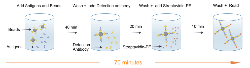
| Assay time | 70 minutes |
| Species reactivity | Human |
Background
Human Interleukin-3 (IL-3), also known as multi-colony stimulating factor (multi-CSF), is a pleiotropic cytokine secreted by T cells and mast cells that regulates hematopoiesis and immunity. Its gene is located at 5q31.1 and is linked to GM-CSF. The receptor consists of an IL-3Rα (CD123) subunit and a shared βc subunit, transducing signals via the JAK2/STAT5 pathway. IL-3 primarily supports the survival, proliferation, and multilineage differentiation of early hematopoietic stem/progenitor cells, including granulocytic, monocytic, erythroid, and megakaryocytic lineages, and is also involved in immune regulation. Clinically, it has been used for chemotherapy-induced myelosuppression and bone marrow failure, and in recent years its applications have expanded to autoimmune fields such as multiple sclerosis and systemic lupus erythematosus. Recombinant human IL-3 is widely used for hematopoietic progenitor cell expansion, TF-1 proliferation assays, and drug development, offering significant research and application value.
Picture
Picture
CBA
Quantifiction of human PBMC
Stimulated sample: Human peripheral blood mononuclear cells (PBMCs) were stimulated with 10 ng/mL PMA+Ionomycin for 24h, and the average concentration of IL-3 in the supernatant was measured to be 2140.05 pg/mL.
Unstimulated sample: Human PBMCs were cultured for 24h, and the average concentration of IL-3 in the supernatant was measured to be 0.2 pg/mL.
Linearity
The high-concentration human IL-3 protein was serially diluted using plasma, serum and cell culture supernatant matrices respectively. Then, the concentration of human IL-3 was determined and interpolated based on the target standard curve, and the sample dilution was corrected.
Recovery
Within the detection range, the combined cytokine proteins were added to different matrices at high,medium and low concentrations.
The matrices used in these experiments were diluted 2-fold with the assay buffer before the addition of the cytokine proteins.
The results were compared with those of samples that had the same concentration of cytokines added in the assay buffer.
Protocol Diagram