Standard Curve
Example of Human IgG4 standard curve in Assay Diluent A1. Plotted are the background-subtracted data.
This standard curve is provided for demonstration only.
Human IgG4 OneStep ELISA Kit
Human IgG4 OneStep ELISA Kit
Price:
Regular price
$120 USD
Regular price
Sale price
$120 USD
Unit price
per
For shipping services or bulk orders, you may request a quotation.
Secure checkout with

View full details
Product Details
Product Details
Product Specification
| Antigen | IgG4 |
| Immunogen | Recombinant Protein |
| Antibody Type | Recombinant mAb |
| Reactivity | Hu |
| Purification | Protein A |
| Stability & Storage | 12 months from date of receipt / reconstitution, 2 to 8°C as supplied. |
Kit
| Precision | Intra-assay: 4.9%; Inter-assay: 5.9% |
| Sample type | Serum; EDTA Plasma, Heparin Plasma, Citrate Plasma; Cell Culture Supernatant |
| Assay type | Sandwich (quantitative) |
| Sensitivity | 113.91 pg/mL |
| Range | 468.8 pg/mL – 30,000 pg/mL |
| Recovery | Serum: 108% EDTA Plasma: 107% Citrate plasma: 95% Heparin Plasma: 100% Cell Culture Supernatant: 103% |
| Assay time | 60 minutes |
| Species reactivity | Hu |
Background
Human IgG4 is the least abundant IgG subclass in serum (1–4% of total IgG) and is structurally and functionally distinct from other subclasses. Its unique hinge and CH2 domain conformation make it largely non-inflammatory, unable to activate complement or bind most Fcγ receptors with high affinity. Its most distinctive feature is Fab-arm exchange, in which half-molecules dissociate and recombine to form bispecific, functionally monovalent antibodies that cannot cross-link identical antigens. Functionally, IgG4 acts as a blocking or tolerance-inducing antibody—protective in allergy and chronic parasitic infection by competing with IgE, but detrimental in cancer by dampening antitumor immunity, and pathogenic in autoimmune diseases such as pemphigus and myasthenia gravis. It is also the hallmark of IgG4-related disease, a fibroinflammatory condition causing tumor-like masses in multiple organs. Due to its reduced effector function, engineered IgG4 (often with the S228P mutation) is a preferred scaffold for therapeutic antibodies where immune activation is undesirable.
Picture
Picture
ELISA
Spike Recovery
The recovery of Human IgG4 was evaluated in activated samples spiked with concentrations spanning the entire assay range.
Dilution Linearity
The concentrations of Human IgG4 were measured and interpolated from the target standard curves and corrected for sample dilution.
The leftmost bar in the graph shows biological samples diluted 10,000-fold in Assay Diluent. "Undiluted" was defined as biological sample diluted 10,000-fold. The mean target concentration was determined to be 108.87 ng/mL in Human plasma-EDTA; The mean target concentration was determined to be 557.83 ng/mL in Human plasma-Heparin; The mean target concentration was determined to be 148.36 ng/mL in Human plasma-Citrate.
Dilution Linearity
The concentrations of Human IgG4 were measured and interpolated from the target standard curves and corrected for sample dilution.
The leftmost bar in the graph shows biological samples diluted 100,000-fold in Assay Diluent. "Undiluted" was defined as biological sample diluted 100,000-fold. The mean target concentration was determined to be 483.07 ng/mL in Human serum.
Spike-and-dilution Linearity
To evaluate assay linearity, three samples were spiked with high levels of Human IgG4 in different matrices and serially diluted with the corresponding Calibrator Diluent to fall within the assay's dynamic range.
Intra-Assay Precision (Precision within an assay)
Three samples of known concentration were tested fifteen times on one plate to assess intra-assay precision.
Inter-Assay Precision (Precision between assays).
Three samples of known concentration were tested in separate assays to assess inter-assay precision. Assays were performed with at least three lots of components.
Determination of Minimum Detectable Dose (MDD)
The MDD was determined using three independent lots of assay components. For each lot, 19 replicate measurements of the diluent (zero calibrator) were performed. The mean (AVERAGE) and standard deviation (STDEV) of the 19 replicates were calculated. The MDD for each lot was then calculated according to the following formula:
MDD = 2 × STDEV + AVERAGE
Cross‑reactivity
The heatmap displays OD450 ratios for serum samples from multiple species. Color intensity is proportional to the O.D.450 ratio (test species / reference species). Darker colors indicate higher cross‑reactivity.
HOOK Effect Threshold
The upper limit of the HOOK effect was established at 100× the highest calibrator concentration (equivalent to a 2-log10 increase). No HOOK effect was observed below this threshold, confirming that samples within this concentration range yield reliable quantitative results without signal depression.
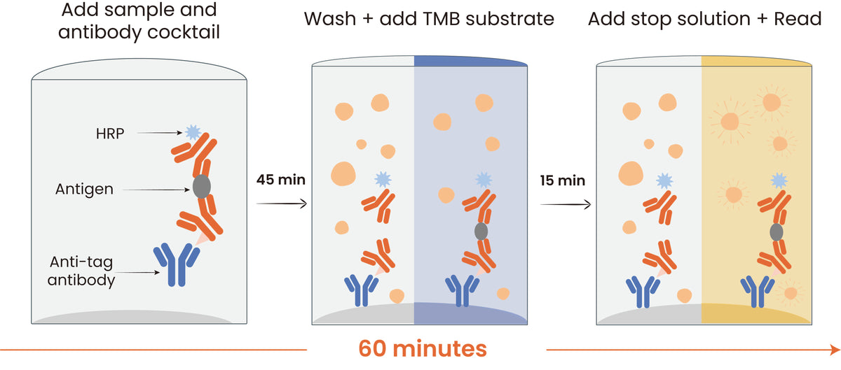
Protocol Diagram