Standard Curve
Example of Human G-CSF standard curve in Assay Diluent A1. Plotted are the background-subtracted data.
This standard curve is provided for demonstration only.
Human G-CSF Culture Media OneStep ELISA
Human G-CSF Culture Media OneStep ELISA
Price:
Regular price
$500 USD
Regular price
Sale price
$500 USD
Unit price
per
For shipping services or bulk orders, you may request a quotation.
Secure checkout with

View full details
Product Details
Product Details
Product Specification
| Antigen | G-CSF |
| Immunogen | Recombinant Protein |
| Antibody Type | Recombinant mAb |
| Reactivity | Hu |
| Purification | Protein A |
| Stability & Storage | 12 months from date of receipt / reconstitution, 2 to 8°C as supplied. |
Kit
| Precision | Intra-assay: 2.7%; Inter-assay: 3.1% |
| Sample type | Cell Culture Supernatant |
| Assay type | Sandwich (quantitative) |
| Sensitivity | 35.30 pg/mL |
| Range | 78.1 pg/mL – 5000 pg/mL |
| Recovery | Cell Culture Supernatant: 98% |
| Assay time | 60 minutes |
| Species reactivity | Hu |
Background
Human Granulocyte Colony-Stimulating Factor (G-CSF) is a ~20 kDa glycoprotein of 174 amino acids that functions as the principal hematopoietic cytokine controlling neutrophil production and function. It is produced mainly by endothelial cells, monocytes-macrophages, and fibroblasts upon stimulation such as infection, and it drives the proliferation, survival, and neutrophilic differentiation of myeloid progenitor cells by activating its specific receptor. This receptor belongs to the hematopoietin receptor superfamily and triggers multiple signaling cascades that balance steady-state and "emergency" granulopoiesis. G-CSF deficiency in mice leads to neutropenia, reduced hematopoietic progenitors, and increased susceptibility to spontaneous infections. Clinically, recombinant human G-CSF is widely used to treat congenital and acquired neutropenias, reduce infectious complications during chemotherapy, and enable dose-intensified cancer treatment. More recent evidence also suggests potential therapeutic applications beyond neutropenia, including cardiac and brain infarction. Thus, human G-CSF serves as both a critical physiological regulator of innate immunity and an essential clinical tool in hematology and oncology.
Picture
Picture
ELISA
Spike Recovery
The recovery of Human G-CSF was evaluated in activated samples spiked with concentrations spanning the entire assay range.
Dilution Linearity
The concentrations of Human G-CSF were measured and interpolated from the target standard curves and corrected for sample dilution.
The leftmost bar in the graph shows biological samples diluted in Assay Diluent to 50% concentration. "undiluted" was defined as 50% biological sample. The subsequent bars represent two-fold serial dilutions of this reference. The mean target concentration was determined to be 955.17 pg/mL in Human Peripheral blood mononuclear cells (PBMCs) stimulated with 10 μg/mL PHA for 5 days. Unstimulated PBMC supernatant signal was undetectable.
Spike-and-dilution Linearity
To evaluate assay linearity, sample was spiked were spiked with high levels of Human G-CSF in different matrices and serially diluted with the corresponding Calibrator Diluent to fall within the assay's dynamic range.
Intra-Assay Precision (Precision within an assay)
Three samples of known concentration were tested fifteen times on one plate to assess intra-assay precision.
Inter-Assay Precision (Precision between assays).
Three samples of known concentration were tested in separate assays to assess inter-assay precision. Assays were performed with at least three lots of components.
Determination of Minimum Detectable Dose (MDD)
The MDD was determined using three independent lots of assay components. For each lot, 19 replicate measurements of the diluent (zero calibrator) were performed. The mean (AVERAGE) and standard deviation (STDEV) of the 19 replicates were calculated. The MDD for each lot was then calculated according to the following formula:
MDD = 2 × STDEV + AVERAGE
Cross‑reactivity
The cross‑reactivity between Human G-CSF and Mouse G-CSF was assessed by testing both proteins in the same assay system. Serial dilutions of each protein were measured, and the dose‑response curves were compared. No significant cross‑reactivity was observed when Mouse G-CSF was tested in the Human G-CSF assay, as the signals remained within the background range of the assay.
HOOK Effect Threshold
The upper limit of the HOOK effect was established at 100× the highest calibrator concentration (equivalent to a 2-log10 increase). No HOOK effect was observed below this threshold, confirming that samples within this concentration range yield reliable quantitative results without signal depression.
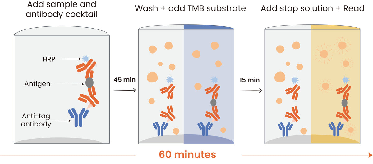
Protocol Diagram