Immunofluorescence detection of characteristic protein expression in vascular organoids: the vascular organoids express endothelial cell marker CD31 (green) and pericyte marker SMA (red).
hPSC Vascular Organoid Differentiation Kit
hPSC Vascular Organoid Differentiation Kit
Price:
Regular price
$1,300 USD
Regular price
Sale price
$1,300 USD
Unit price
per
For shipping services or bulk orders, you may request a quotation.
Secure checkout with

View full details
Product Details
Product Details
Product Specification
| Species | Human |
| Synonyms | hPSC Vascular Organoid Differentiation Kit |
| Stability & Storage | Store according to product components; the reagent kit has a shelf life of 6 months. |
Background
This kit provides a complete solution for inducing the differentiation of human pluripotent stem cells (hPSCs) or human embryonic stem cells (hESCs) into blood vessel organoids (BVOs). The blood vessel organoids are composed of endothelial cells (CD31⁺/VE-Cadherin⁺) and pericytes (PDGFR-β⁺/SMA⁺), which self-assemble in a 3D culture system to form vascular networks with luminal structures. These organoids can be further used for modeling vascular diseases, drug screening, and in vivo transplantation studies.
Using this kit requires that operators have experience in hPSC culture and a certain level of understanding of organoids.
Protocol
Reagents required but not included in this kit
Product Name |
Specifications |
Stem cell culture medium |
500ml |
Stem cell digestion solution |
100 ml |
Matrigel (stem cell culture) |
5 ml |
Ultra-low adsorption6-well plate/24-well plate |
/ |
Extracellular matrix componentsECM1 |
/ |
Extracellular matrix componentsECM2 |
/ |
NaOH |
/ |
HCL |
/ |
DPBS |
/ |
Induced culture of hPSC vascular organoids
Early recoveryhPSC,After at least two generations, cells can be sequentially induced using differentiation media at different stages, with the specific steps as follows:
Embryo body formation (1-2Day)
(1)Cultured in a T25 flaskhPSCTaking cells as an example, when the cell confluence is75–85%, and when most cells are in an undifferentiated state, aspirate the culture medium and add2 mlWash once with DPBS, then aspirate and add 1 ml of Accutase, returning the culture dish to the 37°C incubator until all cells have dissociated into small cell clumps (usually takes 5–7 minutes).
(2) After digestion is complete, add 1 mlof mTeSR™ medium, resuspend the cells into a single-cell suspension, collect the cells and transfer them to a15 ml centrifuge tube, and centrifuge at 300g for 3 minutes at room temperature.
(3)Prepare the embryoid body formation complete medium.
Thaw Vascular Organoid Differentiation Medium A and its Supplement at 4°C in advance. Completely transfer the Supplement into Medium A and mix well to obtain the embryoid body formation complete medium.
Note::For small-volume reagent components, it is recommended to briefly centrifuge for 5 seconds (500–2000 rpm) before opening the cap to avoid loss.
(4)After centrifugation, discard the supernatant and resuspend the cells in embryoid body formation complete medium, adjusting the cell density to3x10⁵-1x10⁶cells/well, toward ultra-low adsorptionSeed 2 per well in a 6-well plate.-3 mL of cell suspension, gently mixed and then incubated in a 37°C incubator.Cultivate 1 in the middle-2Heavens, until a cellular aggregate with smooth boundaries is formed (diameter>50-100 µm), you can proceed to the next step.
Note:Due to aggregation ability inhPSCCell lines may vary; the culture time can be adjusted to 2 as needed.-3Heaven, but the diameter of the embryoid cannot exceed 1.0.00 µm; the excessively large volume impairs subsequent differentiation efficiency.
Mesoderm induction3Day)
(1) Prepare the complete medium for mesoderm induction.
Thaw Vascular Organoid Differentiation Medium B and its Supplement at 4°C in advance. Transfer the Supplement completely into Vascular Organoid Differentiation Medium B and mix thoroughly to obtain the complete mesoderm induction medium.
(2) Use a Pasteur pipette to transfer the embryoid bodies to 1.5Collect by natural sedimentation in an mL centrifuge tube, and remove the supernatant.
(3) Continue to add 2-Resuspend the spheroids in 3 mL of complete mesoderm induction medium and replate them into a 6-well plate, continuing culture for an additional 3 days., inducing mesoderm differentiation.
Note:During the culture period, ensure that the embryoid bodies remain uniformly dispersed. If any aggregates form, gently disperse them using a 1-mL pipette tip with the tip cut off, taking care to avoid excessive fusion.
Vascular lineage induction2Day)
(1)Prepare the complete medium for vascular lineage induction.
Thaw vascular organoid differentiation medium C and its supplement at 4℃ in advance. Completely transfer the supplement into vascular organoid differentiation medium C and mix well to obtain the complete vascular lineage induction medium.
(2)Use a Pasteur pipette to transfer the spheroids into a 15 ml centrifuge tube, allow them to settle naturally, and remove the supernatant.(3) Continue by adding 2 mL of complete vascular lineage induction medium to resuspend the spheroids, then replate them into a 6-well plate and continue culturing for 2 days.
Vascular network induction (9 days)(1)Calculate the volume of angiogenesis matrix required for this stage: angiogenesis is typically performed in a 12-well plate, with 1 mL per well, and each well can accommodate 20–30 spheroids.
Vascular network induction (9 days)
(1)Calculate the volume of angiogenesis matrix needed for this stage: angiogenesis is usually carried out in a 12-well plate, with 1 mL per well, and each well can accommodate 20-30 spheroids.
Note:The density of spheroids inoculated per well is crucial; if it's too dense, the vascular networks may overmerge, while if it's too sparse, the efficiency of vascular network formation will be low.
(2)Prepare the angiogenic matrix on ice. (Using 12 mL as an example.)
Prepare a mixed solution of angiogenesis-specific hydrogel with a pH of 7.4: mix 3.6 mL of EMC1 with 5.4 mL of...Angiogenesis matrix preparation mediumMix to prepare a 9 mL angiogenesis-specific gel mixture, and quickly measure the pH of the mixture using a pH indicator strip; the pH should be approximately 7.4. If the pH does not meet the requirement, adjust it using 1 M NaOH and 1 M HCl.
Preparation of the angiogenesis matrix:Add 9 mLAngiogenesis-specific adhesive mixtureMix thoroughly with 3 mL of EMC2 to obtain the angiogenic matrix.
Note:All operations are performed on ice, and the angiogenesis matrix is prepared and used immediately.
(3) Add 500 μL to each well of a 12-well plate.The angiogenesis matrix was subjected to coating treatment.Incubated at 37℃Allow it to solidify for 1–2 hours.
Note:Avoid the formation of bubbles in the angiogenesis matrix.
(4)Transfer the sphere into 1 using a Pasteur pipette.5 mL centrifuge tube, after natural sedimentation, remove.Remove the supernatant, add 5.mLResuspend in DPBS, allow to settle again, and then remove the supernatant.
(5) Each20-30 spheres in 500 μLResuspend the angiogenesis matrix, according toSeed 500 μL per well into a pre-coated 12-well plate.By shaking the board back and forth, evenly distribute the spheres across its surface.Incubated at 37℃Allow it to solidify for 1–2 hours.
(6) Prepare the complete vascular differentiation medium.
Thaw the vascular organoid differentiation medium D and its additives at 4°C in advance. Completely transfer the additives into the vascular organoid differentiation medium D and mix well to obtain the complete vascular differentiation medium, which can be aliquoted and stored as needed.
(7)Add the complete vascular differentiation medium along the well wall at a volume of1 mL per wellto induce vascular differentiation; vascular buds typically appear within1-3 days.
(8)After culturing for3 days, perform the first medium change, and thereafter change the medium every2–3 days. Continue culturing for another 9 days; approximately10 days later, the vascular organoids can be fixed and stained, or subjected to other protocols for subsequent analysis.
Vascular organoid maturation (2 days)
(1) Separation: Carefully aspirate the upper layer of culture medium from the EMC, separate the EMC from the plate using a sterile blade, and use tools such as forceps to transfer the EMC intact into a 10 cm cell culture dish.
(2) Dissection. Under a stereomicroscope or microscope, use a sterile blade or the tip of a 1 mL syringe to completely dissect individual vascular networks.
Note:Do not disrupt the structure of the vascular network.
(3) Culture. Add 150 μL of fully differentiated vascular organoid culture medium D to an ultra-low attachment 96-well plate, seed one vascular network per well, and incubate in a 37°C cell culture incubator for 2 days.
Note:One standard differentiation batch can support the maturation of at least 200 vascular networks into vascular organoids.
Guidelines
(1) Operations such as product sub-packaging and use must be performed in a sterile environment.
Picture
Picture
Bioactivity
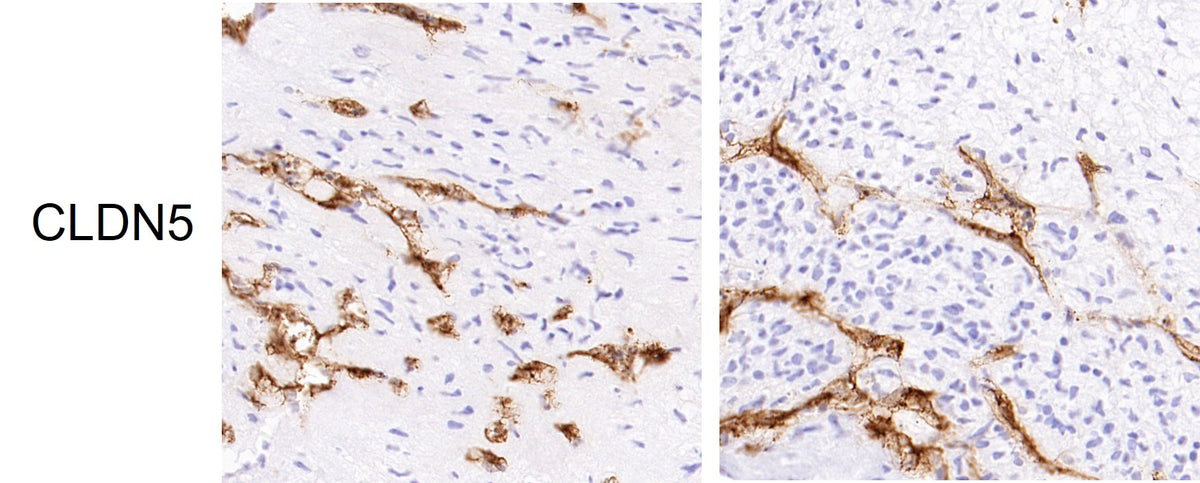
Immunohistochemical identification: the vascular organoids express endothelial cell marker CLDN5 (brown).