WB result of Hormone sensitive lipase/HSL Recombinant Rabbit mAb
Primary antibody: Hormone sensitive lipase/HSL Recombinant Rabbit mAb at 1/1000 dilution
Lane 1: Huh7 whole cell lysate 20 μg
Lane 2: Raji whole cell lysate 20 μg
Negative control: Huh7 whole cell lysate
Secondary antibody: Goat Anti-Rabbit IgG (H+L), HRP conjugated at 1/10000 dilution
Predicted MW: 117 kDa
Observed MW: 100 kDa
Hormone sensitive lipase/HSL Recombinant Rabbit mAb (S-6116)
Hormone sensitive lipase/HSL Recombinant Rabbit mAb (S-6116)
Price:
Regular price
$100 USD
Regular price
Sale price
$100 USD
Unit price
per
For shipping services or bulk orders, you may request a quotation.
Secure checkout with

View full details
Product Details
Product Details
Product Specification
| Host | Rabbit |
| Antigen | Hormone sensitive lipase/HSL |
| Synonyms | Hormone-sensitive lipase; Monoacylglycerol lipase LIPE; Retinyl ester hydrolase (REH); LIPE |
| Location | Cytoplasm, Cell membrane |
| Accession | Q05469 |
| Clone Number | S-6116 |
| Antibody Type | Recombinant mAb |
| Isotype | IgG |
| Application | WB, IHC-P |
| Reactivity | Hu, Ms, Rt |
| Positive Sample | Raji, mouse adipose |
| Purification | Protein A |
| Concentration | 0.5 mg/ml |
| Conjugation | Unconjugated |
| Physical Appearance | Liquid |
| Storage Buffer | PBS, 40% Glycerol, 0.05% BSA, 0.02% sodium azide |
| Stability & Storage | 12 months from date of receipt / reconstitution, -20 °C as supplied |
Dilution
| application | dilution | species |
| WB | 1:1000-1:2000 | Hu, Ms |
| IHC-P | 1:2000 | Hu, Ms, Rt |
Background
Hormone-sensitive lipase (HSL) is an intracellular neutral lipase that was long regarded as the rate-limiting enzyme for triglyceride hydrolysis in adipose tissue; it is now known to act primarily at the diacylglycerol step of the lipolytic cascade, hydrolyzing diacylglycerol to monoacylglycerol. HSL activity is acutely regulated by reversible phosphorylation: catecholamines and other hormones phosphorylate HSL via the cAMP/PKA pathway, promoting its translocation to lipid droplets where it exerts its hydrolytic action, whereas insulin inhibits this process. The enzyme has an unusually broad substrate specificity, hydrolyzing not only triglycerides, diacylglycerols, and monoacylglycerols but also cholesterol esters, steroid fatty acid esters, and retinyl esters. HSL is highly expressed in adipose tissue and steroidogenic tissues, and is also expressed in cardiac muscle, skeletal muscle, macrophages, and pancreatic islets; in steroidogenic tissues it is mainly responsible for converting cholesterol esters into free cholesterol for hormone synthesis. At the pathophysiological level, HSL expression and activity correlate significantly with the maximal lipolytic capacity of human adipocytes, and abnormalities in its activity are involved in obesity, insulin resistance, and the excessive release of free fatty acids in diabetic ketoacidosis.
Picture
Picture
Western Blot
WB result of Hormone sensitive lipase/HSL Recombinant Rabbit mAb
Primary antibody: Hormone sensitive lipase/HSL Recombinant Rabbit mAb at 1/1000 dilution
Lane 1: mouse liver lysate 20 μg
Lane 2: mouse adipose lysate 20 μg
Negative control: mouse liver lysate
Secondary antibody: Goat Anti-Rabbit IgG (H+L), HRP conjugated at 1/10000 dilution
Predicted MW: 117 kDa
Observed MW: 84, 36 kDa
Immunohistochemistry
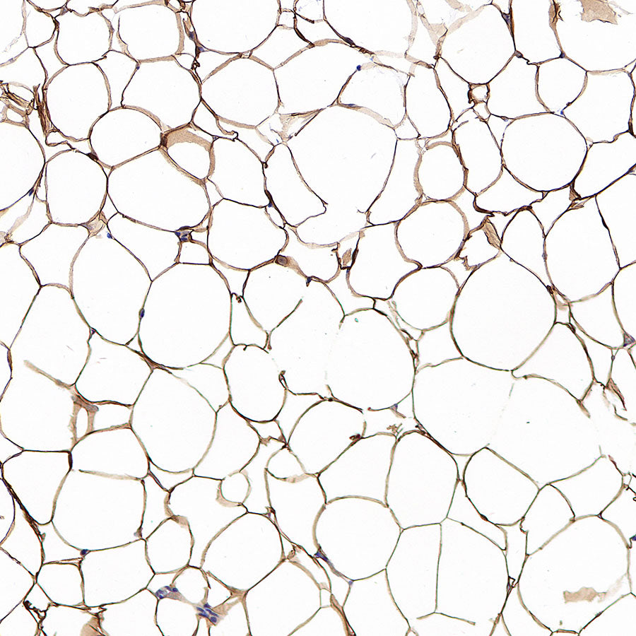
IHC shows positive staining in paraffin-embedded human adipose tissue. Anti-Hormone sensitive lipase/HSL antibody was used at 1/2000 dilution, followed by a HRP Polymer for Rabbit IgG (ready to use). Counterstained with hematoxylin. Heat mediated antigen retrieval with Tris/EDTA buffer pH9.0 was performed before commencing with IHC staining protocol.
Negative control: IHC shows negative staining in paraffin-embedded human liver. Anti- Hormone sensitive lipase/HSL antibody was used at 1/2000 dilution, followed by a HRP Polymer for Rabbit IgG (ready to use). Counterstained with hematoxylin. Heat mediated antigen retrieval with Tris/EDTA buffer pH9.0 was performed before commencing with IHC staining protocol.
IHC shows positive staining in paraffin-embedded mouse adipose tissue. Anti-Hormone sensitive lipase/HSL antibody was used at 1/2000 dilution, followed by a HRP Polymer for Rabbit IgG (ready to use). Counterstained with hematoxylin. Heat mediated antigen retrieval with Tris/EDTA buffer pH9.0 was performed before commencing with IHC staining protocol.
IHC shows positive staining in paraffin-embedded human colon. Anti-Hormone sensitive lipase/HSL antibody was used at 1/2000 dilution, followed by a HRP Polymer for Rabbit IgG (ready to use). Counterstained with hematoxylin. Heat mediated antigen retrieval with Tris/EDTA buffer pH9.0 was performed before commencing with IHC staining protocol.
IHC shows positive staining in paraffin-embedded rat adipose tissue. Anti-Hormone sensitive lipase/HSL antibody was used at 1/2000 dilution, followed by a HRP Polymer for Rabbit IgG (ready to use). Counterstained with hematoxylin. Heat mediated antigen retrieval with Tris/EDTA buffer pH9.0 was performed before commencing with IHC staining protocol.