WB result of HO-1 Rabbit pAb
Primary antibody: HO-1 Rabbit pAb at 1/1000 dilution
Lane 1: untreated HeLa whole cell lysate 20 µg
Lane 2: HeLa treated with 50 µM arsenite for 8 hours whole cell lysate 20 µg
Secondary antibody: Goat Anti- rabbit IgG, (H+L), HRP conjugated at 1/10000 dilution
Predicted MW: 33 kDa
Observed MW: 30 kDa
HO-1 Rabbit Polyclonal Antibody
HO-1 Rabbit Polyclonal Antibody
Price:
Regular price
$100 USD
Regular price
Sale price
$100 USD
Unit price
per
For shipping services or bulk orders, you may request a quotation.
Secure checkout with

View full details
Product Details
Product Details
Product Specification
| Host | Rabbit |
| Antigen | HO-1 |
| Synonyms | Heme oxygenase 1; HO-1Cleaved into 1 chainsHeme oxygenase 1 soluble form; HO; HO1; HMOX1 |
| Immunogen | Recombinant Protein |
| Location | Endoplasmic reticulum |
| Accession | P09601 |
| Antibody Type | Polyclonal antibody |
| Isotype | IgG |
| Application | WB, IHC-P |
| Reactivity | Hu, Ms |
| Positive Sample | HeLa (treated with 50 µM arsenite for 8 hours), NIH/3T3 (treated with 50 µM arsenite for 8 hours) |
| Purification | Immunogen Affinity |
| Concentration | 0.5 mg/ml |
| Conjugation | Unconjugated |
| Physical Appearance | Liquid |
| Storage Buffer | PBS, 40% Glycerol, 0.05% BSA, 0.03% Proclin 300 |
| Stability & Storage | 12 months from date of receipt / reconstitution, -20 °C as supplied |
Dilution
| application | dilution | species |
| WB | 1:1000 | Hu, Ms |
| IHC-P | 1:1000 | Hu, Ms |
Background
Heme Oxygenase-1 (HO-1, HSP32) is a 32 kDa stress-responsive enzyme anchored to the smooth endoplasmic reticulum that cleaves heme at the α-meso carbon to release equimolar biliverdin (rapidly reduced to bilirubin), carbon monoxide (CO) and ferrous iron, thereby not only eliminating the pro-oxidant heme but also generating potent antioxidants and the signaling gas CO; transcription of the HMOX1 gene is potently induced by myriad stressors—including heme itself, ROS, heavy metals, cytokines, hypoxia, nitric oxide, and natural electrophiles—via Nrf2/ARE, Bach1, HIF-1, AP-1 and NF-κB pathways, giving HO-1 a central cytoprotective role against oxidative and inflammatory injury in cardiovascular, neuronal, renal and hepatic tissues, modulating mitochondrial biogenesis, autophagy and immune tolerance, while its products bilirubin and CO respectively scavenge radicals and activate cGMP/PKG signaling to vasodilate, inhibit platelet aggregation and suppress apoptosis, making HO-1 a therapeutic target in sepsis, ischemia–reperfusion, transplant rejection, metabolic disorders and neurodegeneration.
Picture
Picture
Western Blot
WB result of HO-1 Rabbit pAb
Primary antibody: HO-1 Rabbit pAb at 1/1000 dilution
Lane 1: untreated NIH/3T3 whole cell lysate 20 µg
Lane 2: NIH/3T3 treated with 50 µM arsenite for 8 hours whole cell lysate 20 µg
Secondary antibody: Goat Anti- rabbit IgG, (H+L), HRP conjugated at 1/10000 dilution
Predicted MW: 33 kDa
Observed MW: 30 kDa
Immunohistochemistry
IHC shows positive staining in paraffin-embedded human liver. Anti-HO-1 antibody was used at 1/1000 dilution, followed by a HRP Polymer for Rabbit IgG (ready to use). Counterstained with hematoxylin. Heat mediated antigen retrieval with Tris/EDTA buffer pH9.0 was performed before commencing with IHC staining protocol.
IHC shows positive staining in paraffin-embedded human lung. Anti-HO-1 antibody was used at 1/1000 dilution, followed by a HRP Polymer for Rabbit IgG (ready to use). Counterstained with hematoxylin. Heat mediated antigen retrieval with Tris/EDTA buffer pH9.0 was performed before commencing with IHC staining protocol.
IHC shows positive staining in paraffin-embedded human tonsil. Anti-HO-1 antibody was used at 1/1000 dilution, followed by a HRP Polymer for Rabbit IgG (ready to use). Counterstained with hematoxylin. Heat mediated antigen retrieval with Tris/EDTA buffer pH9.0 was performed before commencing with IHC staining protocol.
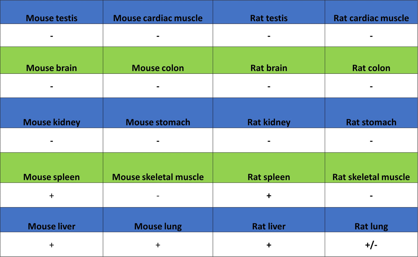
IHC shows positive staining in paraffin-embedded mouse lung. Anti-HO-1 antibody was used at 1/1000 dilution, followed by a HRP Polymer for Rabbit IgG (ready to use). Counterstained with hematoxylin. Heat mediated antigen retrieval with Tris/EDTA buffer pH9.0 was performed before commencing with IHC staining protocol.
IHC shows positive staining in paraffin-embedded mouse spleen. Anti-HO-1 antibody was used at 1/1000 dilution, followed by a HRP Polymer for Rabbit IgG (ready to use). Counterstained with hematoxylin. Heat mediated antigen retrieval with Tris/EDTA buffer pH9.0 was performed before commencing with IHC staining protocol.