WB result of hnRNP A1 Recombinant Rabbit mAb
Primary antibody: hnRNP A1 Recombinant Rabbit mAb at 1/1000 dilution
Lane 1: HeLa whole cell lysate 25 μg
Lane 2: 293T whole cell lysate 25 μg
Lane 3: Hep G2 whole cell lysate 25 μg
Lane 4: U-2 OS whole cell lysate 25 μg
Lane 5: MCF7 whole cell lysate 25 μg
Lane 6: RD whole cell lysate 25 μg
Lane 7: PC-3 whole cell lysate 20 μg
Secondary antibody: Goat Anti-Rabbit IgG (H+L), HRP conjugated at 1/10000 dilution
Predicted MW: 38 kDa
Observed MW: 34 kDa
hnRNP A1 Recombinant Rabbit mAb
hnRNP A1 Recombinant Rabbit mAb
Price:
Regular price
$100 USD
Regular price
Sale price
$100 USD
Unit price
per
For shipping services or bulk orders, you may request a quotation.
Secure checkout with

View full details
Product Details
Product Details
Product Specification
| Host | Rabbit |
| Antigen | hnRNP A1 |
| Location | Cytoplasm, Nucleus |
| Accession | P09651 |
| Antibody Type | Recombinant mAb |
| Isotype | IgG |
| Application | WB, IHC-P |
| Reactivity | Hu, Ms, Rt |
| Positive Sample | HeLa, 293T, HepG2, U-2 OS, MCF7, RD, PC-3, Neuro-2a, C6 |
| Purification | Protein A |
| Concentration | 0.8 mg/ml |
| Conjugation | Unconjugated |
| Physical Appearance | Liquid |
| Storage Buffer | PBS, 40% Glycerol, 0.05% BSA, 0.02% sodium azide |
| Stability & Storage | 12 months from date of receipt / reconstitution, -20 °C as supplied |
Dilution
| application | dilution | species |
| WB | 1:1000-1:2000 | Hu, Ms, Rt |
| IHC-P | 1:100 | Hu |
Background
Heterogeneous nuclear ribonucleoprotein A1 (hnRNP A1) is an abundant RNA-binding protein that serves as a core component of the hnRNP complex, participating in pre-mRNA processing. Its structure consists of two tandem N-terminal RNA recognition motifs (RRM1 and RRM2, collectively known as UP1) and a C-terminal glycine-rich domain, which contains RGG boxes and the M9 nuclear localization signal. hnRNP A1 recognizes YAG motifs in a sequence-specific manner, but its interaction mode with RNA is plastic, allowing it to bind both structured and unstructured RNA. Functionally, hnRNP A1 regulates splice site selection in alternative splicing by antagonizing SR proteins, and also participates in mRNA stability, nuclear export, and translation. Its C-terminal glycine-rich domain mediates protein-protein interactions and confers nucleic acid annealing activity on the protein. In addition, dysregulation of hnRNP A1 is associated with neurodegenerative diseases and viral infections, and disruption of its RNA-binding capacity has become a potential therapeutic target.
Picture
Picture
Western Blot
WB result of hnRNP A1 Recombinant Rabbit mAb
Primary antibody: hnRNP A1 Recombinant Rabbit mAb at 1/1000 dilution
Lane 1: Neuro-2a whole cell lysate 25 μg
Secondary antibody: Goat Anti-Rabbit IgG (H+L), HRP conjugated at 1/10000 dilution
Predicted MW: 38 kDa
Observed MW: 34 kDa
This blot was developed with high sensitivity substrate
WB result of hnRNP A1 Recombinant Rabbit mAb
Primary antibody: hnRNP A1 Recombinant Rabbit mAb at 1/1000 dilution
Lane 1: C6 whole cell lysate 25 μg
Secondary antibody: Goat Anti-Rabbit IgG (H+L), HRP conjugated at 1/10000 dilution
Predicted MW: 38 kDa
Observed MW: 34 kDa
Immunohistochemistry
IHC shows positive staining in paraffin-embedded human colon. Anti-hnRNP A1 antibody was used at 1/100 dilution, followed by a HRP Polymer for Rabbit IgG (ready to use). Counterstained with hematoxylin. Heat mediated antigen retrieval with Tris/EDTA buffer pH9.0 was performed before commencing with IHC staining protocol.
IHC shows positive staining in paraffin-embedded human colon cancer. Anti-hnRNP A1 antibody was used at 1/100 dilution, followed by a HRP Polymer for Rabbit IgG (ready to use). Counterstained with hematoxylin. Heat mediated antigen retrieval with Tris/EDTA buffer pH9.0 was performed before commencing with IHC staining protocol.
IHC shows positive staining in paraffin-embedded human lung cancer. Anti-hnRNP A1 antibody was used at 1/100 dilution, followed by a HRP Polymer for Rabbit IgG (ready to use). Counterstained with hematoxylin. Heat mediated antigen retrieval with Tris/EDTA buffer pH9.0 was performed before commencing with IHC staining protocol.
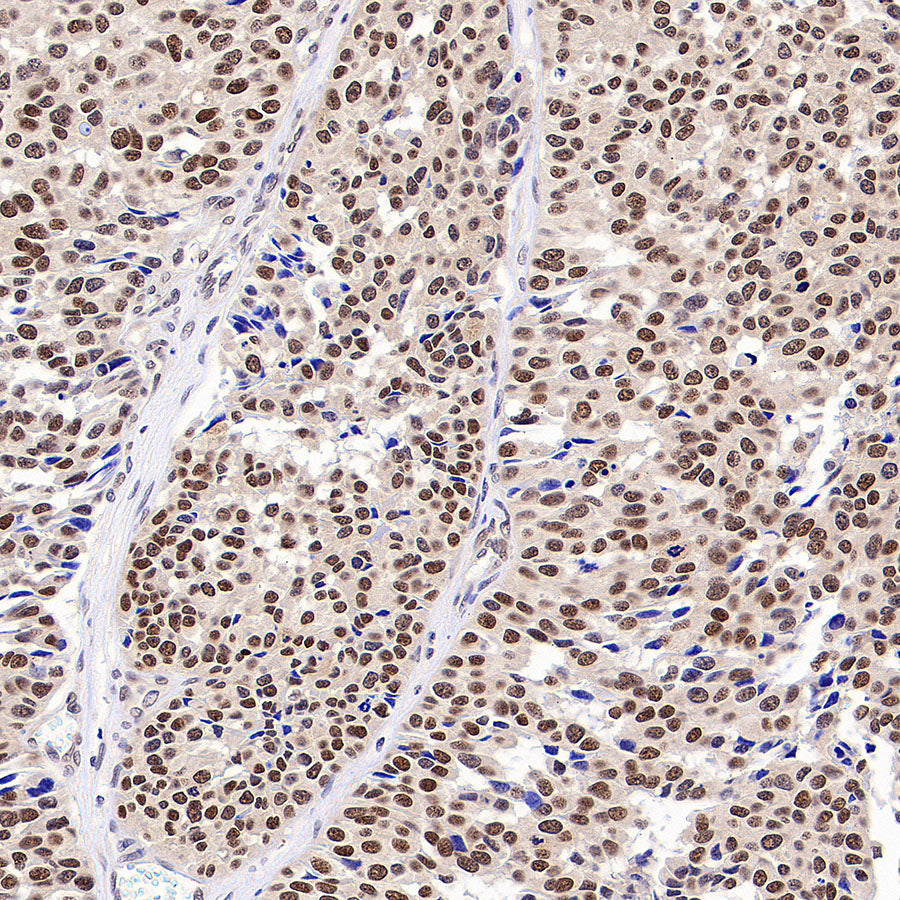
IHC shows positive staining in paraffin-embedded human transitional cell carcinoma. Anti-hnRNP A1 antibody was used at 1/100 dilution, followed by a HRP Polymer for Rabbit IgG (ready to use). Counterstained with hematoxylin. Heat mediated antigen retrieval with Tris/EDTA buffer pH9.0 was performed before commencing with IHC staining protocol.