WB result of His tag Recombinant Rabbit mAb
Primary antibody: His tag Recombinant Rabbit mAb at 1/30000 dilution
Lane 1: 293T transfected with empty vector whole cell lysate 20 µg
Lane 2: 293T transfected with His-tag fusion protein whole cell lysate 20 µg
Secondary antibody: Goat Anti-Rabbit IgG, (H+L), HRP conjugated at 1/10000 dilution
Predicted MW: 60 kDa
Observed MW: 60 kDa
His tag Recombinant Rabbit mAb (S-5315)
His tag Recombinant Rabbit mAb (S-5315)
Price:
Regular price
$100 USD
Regular price
Sale price
$100 USD
Unit price
per
For shipping services or bulk orders, you may request a quotation.
Secure checkout with

View full details
Product Details
Product Details
Product Specification
| Host | Rabbit |
| Antigen | His tag |
| Clone Number | S-5315 |
| Antibody Type | Recombinant mAb |
| Isotype | IgG |
| Application | WB, ICC |
| Reactivity | Species Independent |
| Purification | Protein A |
| Concentration | 0.5 mg/ml |
| Conjugation | Unconjugated |
| Physical Appearance | Liquid |
| Storage Buffer | PBS, 40% Glycerol, 0.05% BSA, 0.02% sodium azide |
| Stability & Storage | 12 months from date of receipt / reconstitution, -20 °C as supplied |
Dilution
| application | dilution | species |
| WB | 1: 5000-1: 50000 | Species independent |
| ICC | 1:200 | Species independent |
Background
A polyhistidine-tag is an amino acid motif in proteins that typically consists of at least six histidine (His) residues, often at the N- or C-terminus of the protein. The total number of histidine residues may vary in the tag from as low as two, to as high as 10 or more His residues.
Picture
Picture
Western Blot
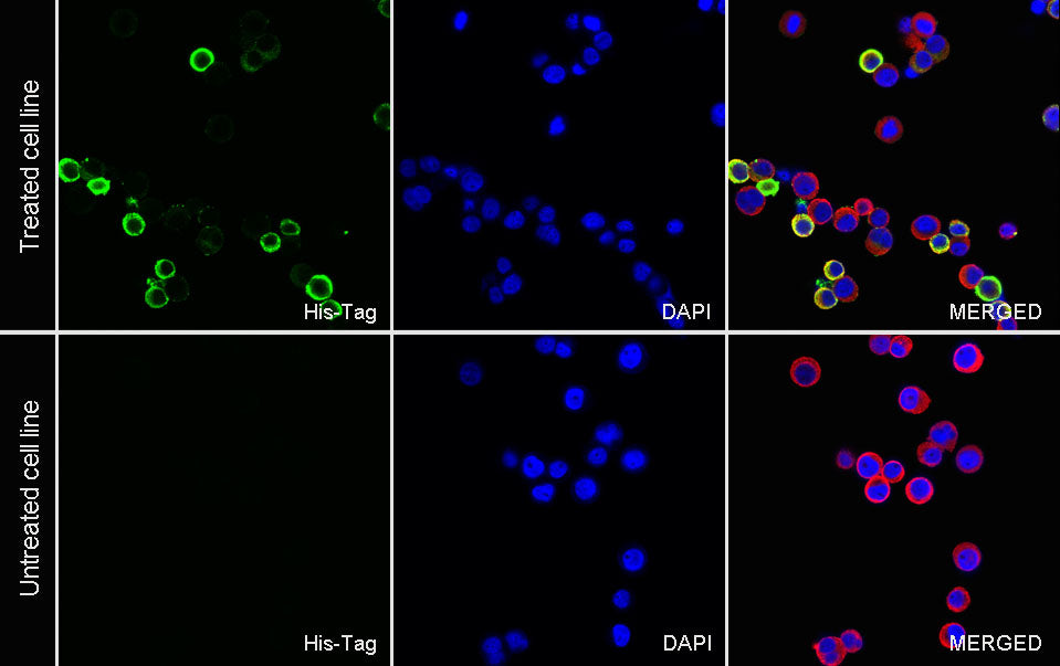
Immunocytochemistry
ICC shows positive staining in MBP tag-his tag transfected 293F cells (top panel) and negative staining in vector-transfected 293F cells (below panel). Anti- His tag antibody was used at 1/200 dilution (Green) and incubated overnight at 4°C. Goat polyclonal Antibody to Rabbit IgG - H&L (Alexa Fluor® 488) was used as secondary antibody at 1/1000 dilution. The cells were fixed with 100% ice-cold methanol and permeabilized with 0.1% PBS-Triton X-100. Nuclei were counterstained with DAPI (Blue). Counterstain with tubulin (Red).