WB result of HIF-1α Recombinant Rabbit mAb
Primary antibody: HIF-1α Recombinant Rabbit mAb at 1/1000 dilution
Primary antibody incubation conditions: overnight at 4°C
Lane 1: untreated Raji whole cell lysate 20 μg
Lane 2: Raji treated with 100 μM chloride for 4 hours whole cell lysate 20 μg
Secondary antibody: Goat Anti-Rabbit IgG (H+L), HRP conjugated at 1/10000 dilution
Predicted MW: 93 kDa
Observed MW: 120 kDa
HIF-1α Recombinant Rabbit mAb (S-6204)
HIF-1α Recombinant Rabbit mAb (S-6204)
Price:
Regular price
$100 USD
Regular price
Sale price
$100 USD
Unit price
per
For shipping services or bulk orders, you may request a quotation.
Secure checkout with

View full details
Product Details
Product Details
Product Specification
| Host | Rabbit |
| Antigen | HIF-1α |
| Synonyms | Hypoxia-inducible factor 1-alpha; HIF-1-alpha; HIF1-alpha; ARNT-interacting protein; Basic-helix-loop-helix-PAS protein MOP1; Class E basic helix-loop-helix protein 78 (bHLHe78); Member of PAS protein 1; PAS domain-containing protein 8; BHLHE78; MOP1; PASD8; HIF1A |
| Location | Cytoplasm, Nucleus |
| Accession | Q16665 |
| Clone Number | S-6204 |
| Antibody Type | Recombinant mAb |
| Isotype | IgG |
| Application | WB, ICC |
| Reactivity | Hu |
| Positive Sample | Raji treated with 100 μM chloride for 4 hours, U-2 OS treated with 1 mM DMOG for 6 hours |
| Predicted Reactivity | Ms, Mk |
| Purification | Protein A |
| Concentration | 0.5 mg/ml |
| Conjugation | Unconjugated |
| Physical Appearance | Liquid |
| Storage Buffer | PBS, 40% Glycerol, 0.05% BSA, 0.02% sodium azide |
| Stability & Storage | 12 months from date of receipt / reconstitution, -20 °C as supplied |
Dilution
| application | dilution | species |
| WB | 1:1000 | Hu |
| ICC | 1:50 | Hu |
Background
HIF-1α (hypoxia-inducible factor-1 alpha) is a crucial transcriptional regulator encoded by the HIF1A gene, playing a vital role in cellular adaptation to low-oxygen conditions. Under normal oxygen levels, HIF-1α has a short half-life and is rapidly degraded via the von Hippel-Lindau tumor suppressor gene product (pVHL)-mediated ubiquitin-proteasome pathway. However, in hypoxic conditions (when oxygen levels are below 5%), HIF-1α becomes stable, accumulates, and translocates to the nucleus. There, it forms an active HIF-1 complex by binding with HIF-1β, which then binds to hypoxia response elements (HRE) in the promoters of target genes, activating the transcription of over 40 genes involved in processes such as oxygen transport, glucose metabolism, cell survival, and angiogenesis. HIF-1α is highly expressed in many tissues, especially in the kidneys and heart, and is often overexpressed in various cancers, contributing to tumor growth and resistance to therapies. Its stability and activity are regulated by various post-translational modifications, including hydroxylation, acetylation, and phosphorylation.
Picture
Picture
Western Blot
WB result of HIF-1α Recombinant Rabbit mAb
Primary antibody: HIF-1α Recombinant Rabbit mAb at 1/1000 dilution
Primary antibody incubation conditions: overnight at 4°C
Lane 1: untreated U-2 OS whole cell lysate 20 μg
Lane 2: U-2 OS treated with 1 mM DMOG for 6 hours whole cell lysate 20 μg
Secondary antibody: Goat Anti-Rabbit IgG (H+L), HRP conjugated at 1/10000 dilution
Predicted MW: 93 kDa
Observed MW: 120 kDa
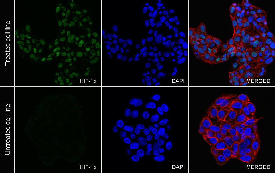
Immunocytochemistry
ICC analysis of HepG2 cells treated with cobalt chloride (500uM,24h) (top panel) and untreated HepG2 cells (below panel). Anti-HIF-1α antibody was used at 1/50 dilution (Green) and incubated overnight at 4°C. Goat polyclonal Antibody to Rabbit IgG - H&L (Alexa Fluor® 488) was used as secondary antibody at 1/1000 dilution. The cells were fixed with 100% ice-cold methanol and permeabilized with 0.1% PBS-Triton X-100. Nuclei were counterstained with DAPI (Blue). Counterstain with tubulin (Red).