WB result of GPRC5A Recombinant Rabbit mAb
Primary antibody: GPRC5A Recombinant Rabbit mAb at 1/1000 dilution
Lane 1: unboiled 293T whole cell lysate 20 µg
Lane 2: unboiled HepG2 whole cell lysate 20 µg
Lane 3: unboiled HeLa whole cell lysate 20 µg
Lane 4: unboiled HT-29 whole cell lysate 20 µg
Lane 5: unboiled MCF7 whole cell lysate 20 µg
Negative control: unboiled 293T whole cell lysate; unboiled HepG2 whole cell lysate
Secondary antibody: Goat Anti- rabbit IgG, (H+L), HRP conjugated at 1/10000 dilution
Predicted MW: 40 kDa
Observed MW: 35-55 kDa
This blot was developed with high sensitivity substrate
GPRC5A Recombinant Rabbit mAb (S-4383-15)
GPRC5A Recombinant Rabbit mAb (S-4383-15)
Price:
Regular price
$100 USD
Regular price
Sale price
$100 USD
Unit price
per
For shipping services or bulk orders, you may request a quotation.
Secure checkout with

View full details
Product Details
Product Details
Product Specification
| Host | Rabbit |
| Antigen | GPRC5A |
| Synonyms | Retinoic acid-induced protein 3; G protein-coupled receptor family C group 5 member A; Phorbol ester induced gene 1 (PEIG-1); Retinoic acid-induced gene 1 protein (RAIG-1); GPCR5A; RAI3; RAIG1 |
| Immunogen | Recombinant Protein |
| Location | Cell membrane |
| Accession | Q8NFJ5 |
| Clone Number | S-4383-15 |
| Antibody Type | Recombinant mAb |
| Isotype | IgG |
| Application | WB, IHC-P, ICC |
| Reactivity | Hu |
| Positive Sample | HeLa, HT-29, MCF7 |
| Purification | Protein A |
| Concentration | 2 mg/ml |
| Conjugation | Unconjugated |
| Physical Appearance | Liquid |
| Storage Buffer | PBS, 40% Glycerol, 0.05% BSA, 0.02% sodium azide |
| Stability & Storage | 12 months from date of receipt / reconstitution, -20 °C as supplied |
Dilution
| application | dilution | species |
| WB | 1:500-1:1000 | Hu |
| IHC-P | 1:200 | Hu |
| ICC | 1:2000 | Hu |
Background
GPRC5A (G protein-coupled receptor class C group 5 member A), also known as retinoic acid-induced gene 1 protein, belongs to the orphan G protein-coupled receptor family. Its structure features the classic seven-transmembrane helix topology, but unlike other GPCRs, it lacks the typical intracellular loop structures required for coupling with heterotrimeric G proteins, suggesting that its signal transduction mechanisms may not depend on classical G protein pathways. Under physiological conditions, GPRC5A is predominantly expressed in epithelial tissues such as the lung, kidney, spleen, and gastrointestinal tract, and its expression is upregulated upon induction by the retinoic acid signaling pathway, participating in the regulation of cell proliferation, differentiation, and tissue development. However, the role of GPRC5A in cancer is exceptionally complex and exhibits a distinct tissue-specific dual function: in lung cancer, GPRC5A is generally considered a tumor suppressor gene—its expression is significantly downregulated in various lung cancer subtypes, and Gprc5a knockout mice spontaneously develop lung adenocarcinoma, indicating that it normally serves as an important barrier against malignant transformation of lung epithelial cells. Conversely, in numerous other malignancies including gastric cancer, esophageal squamous cell carcinoma, colorectal cancer, pancreatic cancer, breast cancer, hepatocellular carcinoma, and glioblastoma, GPRC5A exhibits aberrant overexpression, and its high expression is closely associated with poor patient prognosis, enhanced tumor aggressiveness, epithelial–mesenchymal transition (EMT), and chemoresistance. The underlying pro-tumor mechanisms involve the activation of multiple signaling pathways such as NF-κB, STAT3, and ERK. Given its tumor-specific high expression and functional importance in multiple cancers, GPRC5A is being extensively studied as a promising tumor biomarker and potential therapeutic target. In particular, chimeric antigen receptor T-cell (CAR-T) therapy targeting GPRC5A has demonstrated preliminary antitumor efficacy in preclinical studies against malignancies such as lung cancer.
Picture
Picture
Western Blot
Immunohistochemistry
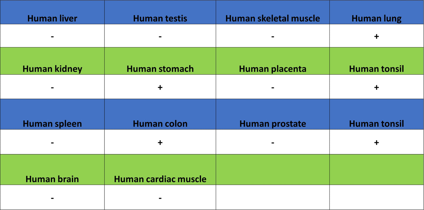
IHC shows positive staining in paraffin-embedded human lung. Anti-GPRC5A antibody was used at 1/200 dilution, followed by a HRP Polymer for Rabbit IgG (ready to use). Counterstained with hematoxylin. Heat mediated antigen retrieval with Tris/EDTA buffer pH9.0 was performed before commencing with IHC staining protocol.
IHC shows positive staining in paraffin-embedded human breast cancer. Anti-GPRC5A antibody was used at 1/200 dilution, followed by a HRP Polymer for Rabbit IgG (ready to use). Counterstained with hematoxylin. Heat mediated antigen retrieval with Tris/EDTA buffer pH9.0 was performed before commencing with IHC staining protocol.
IHC shows positive staining in paraffin-embedded human colon cancer. Anti-GPRC5A antibody was used at 1/200 dilution, followed by a HRP Polymer for Rabbit IgG (ready to use). Counterstained with hematoxylin. Heat mediated antigen retrieval with Tris/EDTA buffer pH9.0 was performed before commencing with IHC staining protocol.
IHC shows positive staining in paraffin-embedded human lung cancer. Anti-GPRC5A antibody was used at 1/200 dilution, followed by a HRP Polymer for Rabbit IgG (ready to use). Counterstained with hematoxylin. Heat mediated antigen retrieval with Tris/EDTA buffer pH9.0 was performed before commencing with IHC staining protocol.
Negative control: IHC shows negative staining in paraffin-embedded human skeletal muscle. Anti-GPRC5A antibody was used at 1/200 dilution, followed by a HRP Polymer for Rabbit IgG (ready to use). Counterstained with hematoxylin. Heat mediated antigen retrieval with Tris/EDTA buffer pH9.0 was performed before commencing with IHC staining protocol.
Immunocytochemistry
ICC shows positive staining in HT-29 cells (top panel) and negative staining in 293T cells (below panel). Anti-GPRC5A antibody was used at 1/2000 dilution (Green) and incubated overnight at 4°C. Goat polyclonal Antibody to Rabbit IgG - H&L (Alexa Fluor® 488) was used as secondary antibody at 1/1000 dilution. The cells were fixed with 100% ice-cold methanol and permeabilized with 0.1% PBS-Triton X-100. Nuclei were counterstained with DAPI (Blue). Counterstain with tubulin (Red).