WB result of GPR153 Recombinant Rabbit mAb
Primary antibody: GPR153 Recombinant Rabbit mAb at 1/1000 dilution
Lane 1: Jurkat whole cell lysate 20 µg
Lane 2: K-562 whole cell lysate 20 µg
Lane 3: A549 whole cell lysate 20 µg
Secondary antibody: Goat Anti-Rabbit IgG (H+L), HRP conjugated at 1/10000 dilution
Predicted MW: 65 kDa
Observed MW: 60 kDa
This blot was developed with high sensitivity substrate
GPR153 Recombinant Rabbit mAb (S-4871-4)
GPR153 Recombinant Rabbit mAb (S-4871-4)
Price:
Regular price
$100 USD
Regular price
Sale price
$100 USD
Unit price
per
For shipping services or bulk orders, you may request a quotation.
Secure checkout with

View full details
Product Details
Product Details
Product Specification
| Host | Rabbit |
| Antigen | GPR153 |
| Synonyms | Probable G protein-coupled receptor 153; PGR1 |
| Immunogen | Synthetic Peptide |
| Location | Cell membrane |
| Accession | Q6NV75 |
| Clone Number | S-4871-4 |
| Antibody Type | Recombinant mAb |
| Isotype | IgG |
| Application | WB, IHC-P |
| Reactivity | Hu, Ms, Mk |
| Positive Sample | Jurkat, K-562, A549, mouse lung, COS7 |
| Purification | Protein A |
| Concentration | 2 mg/ml |
| Conjugation | Unconjugated |
| Physical Appearance | Liquid |
| Storage Buffer | PBS, 40% Glycerol, 0.05% BSA, 0.02% sodium azide |
| Stability & Storage | 12 months from date of receipt / reconstitution, -20 °C as supplied |
Dilution
| application | dilution | species |
| WB | 1:1000 | Hu, Ms, Mk |
| IHC-P | 1:400 | Hu |
Background
GPR153 is an orphan receptor whose endogenous ligand and specific functions remain largely unknown, belonging to the rhodopsin-like class A family of G protein-coupled receptors (GPCRs). The protein features a typical seven-transmembrane α-helical structure, with three extracellular and three intracellular loops connecting the helices, and its predicted ligand-binding pocket is located in the core region formed by the transmembrane helices. A notable structural feature is that GPR153 substitutes the highly conserved DRY motif—essential for efficient G protein signaling in classical class A receptors—with an HRM motif in the third transmembrane helix (TM3), and contains an LPxFL motif (a variant of the NPxxY motif) at the end of TM7; this unique architecture may confer atypical signaling mechanisms. GPR153 shares a common evolutionary origin with GPR162 and is highly expressed in the central nervous system, particularly in the thalamus, cerebellum, and the arcuate nucleus of the hypothalamus—regions closely associated with sensory information processing, motor coordination, and the regulation of energy balance and feeding behavior. Studies have shown that it is also expressed in the mouse neocortex, striatum, substantia nigra, and other regions, suggesting a potential role in broader physiological processes such as decision-making and reward circuits. Furthermore, GPR153 expression has been detected in mouse ovaries, mammary glands, and various other tissues, and its protein has been localized to the cytoplasm and processes of astrocytes in the human brain; however, its precise physiological role in the central nervous system remains an active area of current research.
Picture
Picture
Western Blot
WB result of GPR153 Recombinant Rabbit mAb
Primary antibody: GPR153 Recombinant Rabbit mAb at 1/1000 dilution
Lane 1: mouse lung lysate 20 µg
Secondary antibody: Goat Anti-Rabbit IgG (H+L), HRP conjugated at 1/10000 dilution
Predicted MW: 65 kDa
Observed MW: 60 kDa
This blot was developed with high sensitivity substrate
WB result of GPR153 Recombinant Rabbit mAb
Primary antibody: GPR153 Recombinant Rabbit mAb at 1/1000 dilution
Lane 1: COS7 whole cell lysate 20 µg
Secondary antibody: Goat Anti-Rabbit IgG (H+L), HRP conjugated at 1/10000 dilution
Predicted MW: 65 kDa
Observed MW: 60 kDa
This blot was developed with high sensitivity substrate
Immunohistochemistry
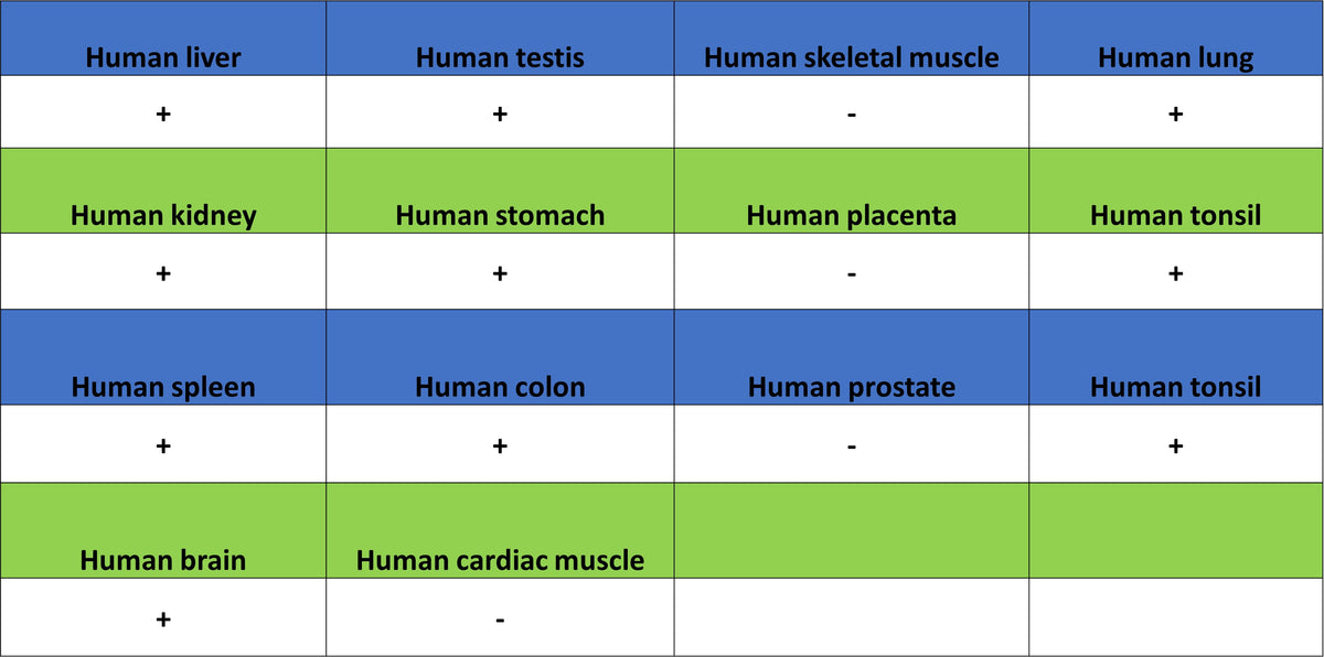
IHC shows positive staining in paraffin-embedded human cerebral cortex. Anti-GPR153 antibody was used at 1/400 dilution, followed by a HRP Polymer for Rabbit IgG (ready to use). Counterstained with hematoxylin. Heat mediated antigen retrieval with Tris/EDTA buffer pH9.0 was performed before commencing with IHC staining protocol.
IHC shows positive staining in paraffin-embedded human liver. Anti-GPR153 antibody was used at 1/400 dilution, followed by a HRP Polymer for Rabbit IgG (ready to use). Counterstained with hematoxylin. Heat mediated antigen retrieval with Tris/EDTA buffer pH9.0 was performed before commencing with IHC staining protocol.