WB result of GOT2 Recombinant Rabbit mAb
Primary antibody: GOT2 Recombinant Rabbit mAb at 1/1000 dilution
Lane 1: Hep G2 whole cell lysate 25 μg
Lane 2: K-562 whole cell lysate 25 μg
Lane 3: SH-SY5Y whole cell lysate 25 μg
Lane 4: A431 whole cell lysate 25 μg
Lane 5: HEK-293 whole cell lysate 25 μg
Lane 6: HeLa whole cell lysate 25 μg
Lane 7: PC-3 whole cell lysate 25 μg
Secondary antibody: Goat Anti-Rabbit IgG (H+L), HRP conjugated at 1/10000 dilution
Predicted MW: 48 kDa
Observed MW: 39 kDa
GOT2 Recombinant Rabbit mAb
GOT2 Recombinant Rabbit mAb
Price:
Regular price
$100 USD
Regular price
Sale price
$100 USD
Unit price
per
For shipping services or bulk orders, you may request a quotation.
Secure checkout with

View full details
Product Details
Product Details
Product Specification
| Host | Rabbit |
| Antigen | GOT2 |
| Location | Mitochondrion, Cell membrane |
| Accession | P00505 |
| Antibody Type | Recombinant mAb |
| Isotype | IgG |
| Application | WB, IHC-P |
| Reactivity | Hu, Ms, Rt |
| Positive Sample | Hep G2, K-562, SH-SY5Y, A431, HEK-293, HeLa, PC-3, mouse brain, mouse kidney, mouse liver, rat brain, rat kidney |
| Purification | Protein A |
| Concentration | 1 mg/ml |
| Conjugation | Unconjugated |
| Physical Appearance | Liquid |
| Storage Buffer | PBS, 40% Glycerol, 0.05% BSA, 0.02% sodium azide |
| Stability & Storage | 12 months from date of receipt / reconstitution, -20 °C as supplied |
Dilution
| application | dilution | species |
| WB | 1:1000-1:5000 | Hu, Ms, Rt |
| IHC-P | 1:100-1:400 | Hu, Ms, Rt |
Background
GOT2 (glutamate-oxaloacetate transaminase 2), also known as mitochondrial aspartate aminotransferase (mAspAT), is a pyridoxal phosphate-dependent enzyme encoded by the GOT2 gene, localized to the inner mitochondrial membrane and matrix. As a key component of the malate-aspartate shuttle system, GOT2 catalyzes the reversible transamination between aspartate and α-ketoglutarate to generate oxaloacetate and glutamate, playing a central role in maintaining NAD(H) redox balance and metabolite exchange between the cytosol and mitochondria. This protein also possesses multiple additional functions: it can catalyze the transamination of L-kynurenine to kynurenic acid and acts as a fatty acid-binding protein promoting cellular uptake of long-chain free fatty acids. GOT2 is highly expressed in the heart and muscle, and its dysregulated expression is associated with various diseases: it is downregulated in clear cell renal cell carcinoma and correlated with poor prognosis, its low expression in hepatocellular carcinoma promotes tumor progression and glutamine metabolic reprogramming, and in cutaneous melanoma it promotes tumor cell proliferation. In addition, GOT2 is regulated by STUB1-mediated ubiquitination and degradation and is involved in the pathogenesis of developmental and epileptic encephalopathy 82.
Picture
Picture
Western Blot
WB result of GOT2 Recombinant Rabbit mAb
Primary antibody: GOT2 Recombinant Rabbit mAb at 1/1000 dilution
Lane 1: mouse brain lysate 25 μg
Lane 2: mouse kidney lysate 25 μg
Lane 3: mouse liver lysate 25 μg
Secondary antibody: Goat Anti-Rabbit IgG (H+L), HRP conjugated at 1/10000 dilution
Predicted MW: 48 kDa
Observed MW: 39 kDa
WB result of GOT2 Recombinant Rabbit mAb
Primary antibody: GOT2 Recombinant Rabbit mAb at 1/1000 dilution
Lane 1: rat brain lysate 25 μg
Lane 2: rat kidney lysate 25 μg
Secondary antibody: Goat Anti-Rabbit IgG (H+L), HRP conjugated at 1/10000 dilution
Predicted MW: 48 kDa
Observed MW: 39 kDa
Immunohistochemistry
IHC shows positive staining in paraffin-embedded human kidney. Anti-GOT2 antibody was used at 1/100 dilution, followed by a HRP Polymer for Rabbit IgG (ready to use). Counterstained with hematoxylin. Heat mediated antigen retrieval with Tris/EDTA buffer pH9.0 was performed before commencing with IHC staining protocol.
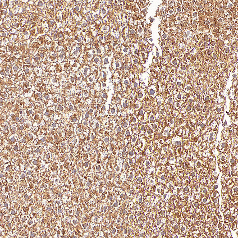
IHC shows positive staining in paraffin-embedded human liver. Anti-GOT2 antibody was used at 1/100 dilution, followed by a HRP Polymer for Rabbit IgG (ready to use). Counterstained with hematoxylin. Heat mediated antigen retrieval with Tris/EDTA buffer pH9.0 was performed before commencing with IHC staining protocol.
IHC shows positive staining in paraffin-embedded human cardiac muscle. Anti-GOT2 antibody was used at 1/100 dilution, followed by a HRP Polymer for Rabbit IgG (ready to use). Counterstained with hematoxylin. Heat mediated antigen retrieval with Tris/EDTA buffer pH9.0 was performed before commencing with IHC staining protocol.
IHC shows positive staining in paraffin-embedded human liver cancer. Anti-GOT2 antibody was used at 1/100 dilution, followed by a HRP Polymer for Rabbit IgG (ready to use). Counterstained with hematoxylin. Heat mediated antigen retrieval with Tris/EDTA buffer pH9.0 was performed before commencing with IHC staining protocol.
IHC shows positive staining in paraffin-embedded human ovarian cancer. Anti-GOT2 antibody was used at 1/100 dilution, followed by a HRP Polymer for Rabbit IgG (ready to use). Counterstained with hematoxylin. Heat mediated antigen retrieval with Tris/EDTA buffer pH9.0 was performed before commencing with IHC staining protocol.
IHC shows positive staining in paraffin-embedded mouse brain. Anti-GOT2 antibody was used at 1/100 dilution, followed by a HRP Polymer for Rabbit IgG (ready to use). Counterstained with hematoxylin. Heat mediated antigen retrieval with Tris/EDTA buffer pH9.0 was performed before commencing with IHC staining protocol.
IHC shows positive staining in paraffin-embedded mouse kidney. Anti-GOT2 antibody was used at 1/100 dilution, followed by a HRP Polymer for Rabbit IgG (ready to use). Counterstained with hematoxylin. Heat mediated antigen retrieval with Tris/EDTA buffer pH9.0 was performed before commencing with IHC staining protocol.
IHC shows positive staining in paraffin-embedded rat kidney. Anti-GOT2 antibody was used at 1/100 dilution, followed by a HRP Polymer for Rabbit IgG (ready to use). Counterstained with hematoxylin. Heat mediated antigen retrieval with Tris/EDTA buffer pH9.0 was performed before commencing with IHC staining protocol.
IHC shows positive staining in paraffin-embedded rat liver. Anti-GOT2 antibody was used at 1/100 dilution, followed by a HRP Polymer for Rabbit IgG (ready to use). Counterstained with hematoxylin. Heat mediated antigen retrieval with Tris/EDTA buffer pH9.0 was performed before commencing with IHC staining protocol.