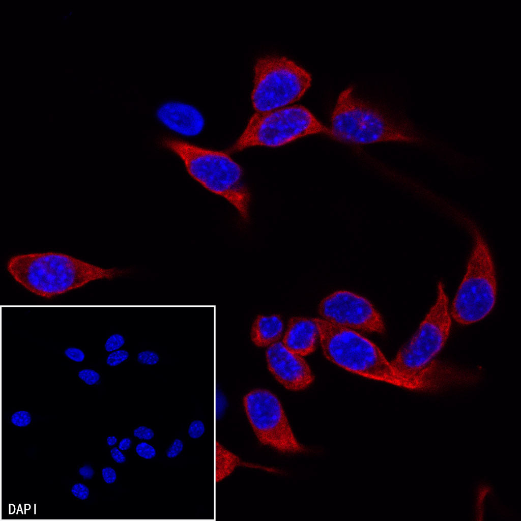
ICC shows positive staining in NIH/3T3 cells. Anti- α-tubulin antibody (S0B0645) was used at 1/1000 dilution (Red) and incubated overnight at 4°C. Goat anti-Mouse IgG(H+L) (TRITC Conjugate) was used as secondary antibody at 1/1000 dilution. The cells were fixed with 100% ice-cold methanol and permeabilized with 0.1% PBS-Triton X-100. Nuclei were counterstained with DAPI (Blue).
Goat anti-Mouse IgG(H+L) (TRITC Conjugate)
Goat anti-Mouse IgG(H+L) (TRITC Conjugate)
Price:
Regular price
$150 USD
Regular price
Sale price
$150 USD
Unit price
per
For shipping services or bulk orders, you may request a quotation.
Secure checkout with

View full details
Product Details
Product Details
Product Specification
| Host | Goat |
| Antigen | Mouse IgG |
| Antibody Type | Polyclonal antibody |
| Application | ICC |
| Reactivity | Ms |
| Purification | Immunogen Affinity |
| Concentration | 2 mg/ml |
| Conjugation | TRITC |
| Physical Appearance | Liquid |
| Storage Buffer | PBS, 1% BSA, 0.09% sodium azid |
| Stability & Storage | 12 months from date of receipt / reconstitution, 2 to 8 °C as supplied |
Dilution
| application | dilution | species |
| ICC | 1:1000 | Ms |
Picture
Picture
Immunocytochemistry
ICC shows positive staining in NIH/3T3 cells. Anti- α-tubulin antibody (S0B0645) was used at 1/1000 dilution (Red) and incubated overnight at 4°C. Goat anti-Mouse IgG(H+L) (TRITC Conjugate) was used as secondary antibody at 1/1000 dilution. The cells were fixed with 4% PFA and permeabilized with 0.1% PBS-Triton X-100. Nuclei were counterstained with DAPI (Blue).