WB result of Glypican-3 Recombinant Rabbit mAb
Primary antibody: Glypican-3 Recombinant Rabbit mAb at 1/1000 dilution
Lane 1: Raji whole cell lysate 20 µg
Lane 2: HepG2 whole cell lysate 20 µg
Negative control: Raji whole cell lysate
Secondary antibody: Goat Anti- rabbit IgG, (H+L), HRP conjugated at 1/10000 dilution
Predicted MW: 65 kDa
Observed MW: 70 kDa
Glypican-3 Recombinant Rabbit mAb (SDT-4064-69-2)
Glypican-3 Recombinant Rabbit mAb (SDT-4064-69-2)
Price:
Regular price
$100 USD
Regular price
Sale price
$100 USD
Unit price
per
For shipping services or bulk orders, you may request a quotation.
Secure checkout with

View full details
Product Details
Product Details
Product Specification
| Host | Rabbit |
| Antigen | Glypican-3 |
| Synonyms | GTR2-2; Intestinal protein OCI-5; MXR7; OCI5; GPC3 |
| Immunogen | Synthetic Peptide |
| Location | Cell membrane |
| Accession | P51654 |
| Clone Number | SDT-4064-69-2 |
| Antibody Type | Recombinant mAb |
| Isotype | IgG |
| Application | WB, IHC-P, ICC |
| Reactivity | Hu |
| Positive Sample | HepG2 |
| Purification | Protein A |
| Concentration | 0.5 mg/ml |
| Conjugation | Unconjugated |
| Physical Appearance | Liquid |
| Storage Buffer | PBS, 40% Glycerol, 0.05% BSA, 0.03% Proclin 300 |
| Stability & Storage | 12 months from date of receipt / reconstitution, -20 °C as supplied |
Dilution
| application | dilution | species |
| WB | 1:1000-1:2000 | Hu |
| IHC-P | 1:500 | Hu |
| ICC | 1:500 | Hu |
Background
Glypican-3 (GPC3) is a cell-surface glycoprotein that is highly expressed in hepatocellular carcinoma (HCC) and some other cancers. It is a member of the glypican family of heparan sulfate proteoglycans, consisting of a core protein and heparan sulfate chains, and is anchored to the cell membrane via a glycosylphosphatidylinositol (GPI) anchor. GPC3 is encoded by the GPC3 gene located on the X chromosome and is cleaved by furin into an N-terminal 40 kDa subunit and a C-terminal 30 kDa subunit. It plays a role in cell proliferation, morphogenesis, and oncogenesis by interacting with signaling pathways such as Wnt, Hedgehog, and fibroblast growth factor. Abnormal expression of GPC3 is associated with poor prognosis in cancer, making it a potential target for cancer diagnosis and immunotherapy.
Picture
Picture
Western Blot
Immunohistochemistry
IHC shows positive staining in paraffin-embedded human placenta. Anti- Glypican-3 antibody was used at 1/500 dilution, followed by a HRP Polymer for Rabbit IgG (ready to use). Counterstained with hematoxylin. Heat mediated antigen retrieval with Tris/EDTA buffer pH9.0 was performed before commencing with IHC staining protocol.
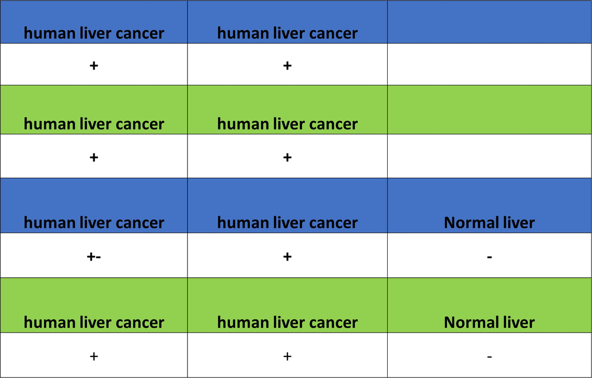
IHC shows positive staining in paraffin-embedded human liver cancer (case 1). Anti- Glypican-3 antibody was used at 1/500 dilution, followed by a HRP Polymer for Rabbit IgG (ready to use). Counterstained with hematoxylin. Heat mediated antigen retrieval with Tris/EDTA buffer pH9.0 was performed before commencing with IHC staining protocol.
IHC shows positive staining in paraffin-embedded human liver cancer (case 2). Anti- Glypican-3 antibody was used at 1/500 dilution, followed by a HRP Polymer for Rabbit IgG (ready to use). Counterstained with hematoxylin. Heat mediated antigen retrieval with Tris/EDTA buffer pH9.0 was performed before commencing with IHC staining protocol.
IHC shows positive staining in paraffin-embedded human liver cancer (case 3). Anti- Glypican-3 antibody was used at 1/500 dilution, followed by a HRP Polymer for Rabbit IgG (ready to use). Counterstained with hematoxylin. Heat mediated antigen retrieval with Tris/EDTA buffer pH9.0 was performed before commencing with IHC staining protocol.
Negative control: IHC shows negative staining in paraffin-embedded human cardiac muscle. Anti-Glypican-3 antibody was used at 1/500 dilution, followed by a HRP Polymer for Rabbit IgG (ready to use). Counterstained with hematoxylin. Heat mediated antigen retrieval with Tris/EDTA buffer pH9.0 was performed before commencing with IHC staining protocol.
Negative control: IHC shows negative staining in paraffin-embedded human kidney. Anti-Glypican-3 antibody was used at 1/500 dilution, followed by a HRP Polymer for Rabbit IgG (ready to use). Counterstained with hematoxylin. Heat mediated antigen retrieval with Tris/EDTA buffer pH9.0 was performed before commencing with IHC staining protocol.
Negative control: IHC shows negative staining in paraffin-embedded human liver. Anti-Glypican-3 antibody was used at 1/500 dilution, followed by a HRP Polymer for Rabbit IgG (ready to use). Counterstained with hematoxylin. Heat mediated antigen retrieval with Tris/EDTA buffer pH9.0 was performed before commencing with IHC staining protocol.
Negative control: IHC shows negative staining in paraffin-embedded human liver. Anti-Glypican-3 antibody was used at 1/500 dilution, followed by a HRP Polymer for Rabbit IgG (ready to use). Counterstained with hematoxylin. Heat mediated antigen retrieval with Tris/EDTA buffer pH9.0 was performed before commencing with IHC staining protocol.
Immunocytochemistry
ICC shows positive staining in HepG2 cells (top panel) and negative staining in Raji cells (below panel). Anti-Glypican-3 antibody was used at 1/500 dilution (Green) and incubated overnight at 4°C. Goat polyclonal Antibody to Rabbit IgG - H&L (Alexa Fluor® 488) was used as secondary antibody at 1/1000 dilution. The cells were fixed with 100% ice-cold methanol and permeabilized with 0.1% PBS-Triton X-100. Nuclei were counterstained with DAPI (Blue). Counterstain with tubulin (Red).