WB result of GLUT3 + GLUT14 Recombinant Rabbit mAb
Primary antibody: GLUT3 + GLUT14 Recombinant Rabbit mAb at 1/1000 dilution
Lane 1: SW480 whole cell lysate 20 µg
Lane 2: HepG2 whole cell lysate 20 µg
Secondary antibody: Goat Anti-Rabbit IgG (H+L), HRP conjugated at 1/10000 dilution
Predicted MW: 54, 55 kDa
Observed MW: 40-60 kDa
GLUT3 + GLUT14 Recombinant Rabbit mAb (S-5945)
GLUT3 + GLUT14 Recombinant Rabbit mAb (S-5945)
Price:
Regular price
$100 USD
Regular price
Sale price
$100 USD
Unit price
per
For shipping services or bulk orders, you may request a quotation.
Secure checkout with

View full details
Product Details
Product Details
Product Specification
| Host | Rabbit |
| Antigen | GLUT3 + GLUT14 |
| Location | Cell membrane |
| Accession | P11169 |
| Clone Number | S-5945 |
| Antibody Type | Recombinant mAb |
| Isotype | IgG |
| Application | WB, IHC-P |
| Reactivity | Hu, Ms, Rt |
| Positive Sample | SW480, HepG2, mouse testis, rat testis |
| Purification | Protein A |
| Concentration | 0.5 mg/ml |
| Conjugation | Unconjugated |
| Physical Appearance | Liquid |
| Storage Buffer | PBS, 40% Glycerol, 0.05% BSA, 0.02% sodium azide |
| Stability & Storage | 12 months from date of receipt / reconstitution, -20 °C as supplied |
Dilution
| application | dilution | species |
| WB | 1:1000-1:5000 | Hu, Ms, Rt |
| IHC-P | 1:2000 | Hu, Ms, Rt |
Background
GLUT3 (SLC2A3) is a member of the Class I facilitative glucose transporter family, characterized by high affinity and high transport capacity, and is mainly expressed in neurons, sperm, and embryonic tissues. Its Km value is approximately 1.4 mM, and its transport rate (Kcat ≈ 6,500/s) is significantly higher than that of GLUT1 and GLUT4, enabling neurons to efficiently take up glucose even in the low-glucose environment of the brain interstitium, where glucose concentrations are only 1–2 mM. GLUT14 (SLC2A14) is a gene duplication product of GLUT3, sharing 94.5% amino acid sequence homology with it, but GLUT14 is mainly expressed in the testis, where its mRNA level is approximately four times that of GLUT3, while it is almost not expressed in brain tissue. GLUT14 has two splice variants, a long form (520 aa) and a short form (497 aa), differing only at the N-terminus, and its functional substrate profile has not been fully elucidated, leading it to be regarded as an "orphan transporter."
Picture
Picture
Western Blot
WB result of GLUT3 + GLUT14 Recombinant Rabbit mAb
Primary antibody: GLUT3 + GLUT14 Recombinant Rabbit mAb at 1/1000 dilution
Lane 1: mouse testis lysate 20 µg
Secondary antibody: Goat Anti-Rabbit IgG (H+L), HRP conjugated at 1/10000 dilution
Predicted MW: 54, 55 kDa
Observed MW: 38-40 kDa
WB result of GLUT3 + GLUT14 Recombinant Rabbit mAb
Primary antibody: GLUT3 + GLUT14 Recombinant Rabbit mAb at 1/1000 dilution
Lane 1: rat testis lysate 20 µg
Secondary antibody: Goat Anti-Rabbit IgG (H+L), HRP conjugated at 1/10000 dilution
Predicted MW: 54, 55 kDa
Observed MW: 38-42 kDa
Immunohistochemistry
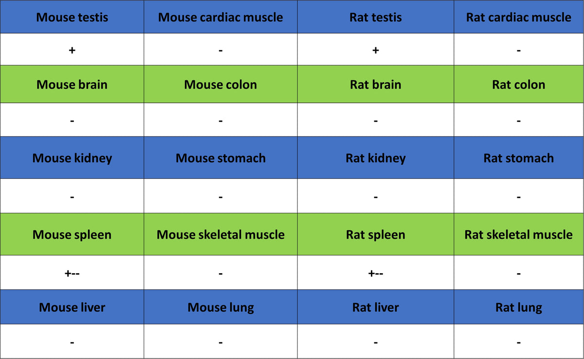
IHC shows positive staining in paraffin-embedded human brain. Anti-GLUT3+GLUT4 antibody was used at 1/2000 dilution, followed by a HRP Polymer for Rabbit IgG (ready to use). Counterstained with hematoxylin. Heat mediated antigen retrieval with Tris/EDTA buffer pH9.0 was performed before commencing with IHC staining protocol.
IHC shows positive staining in paraffin-embedded human testis. Anti-GLUT3+GLUT4 antibody was used at 1/2000 dilution, followed by a HRP Polymer for Rabbit IgG (ready to use). Counterstained with hematoxylin. Heat mediated antigen retrieval with Tris/EDTA buffer pH9.0 was performed before commencing with IHC staining protocol.
Negative control: IHC shows negative staining in paraffin-embedded human skeletal muscle. Anti-GLUT3+GLUT4 antibody was used at 1/2000 dilution, followed by a HRP Polymer for Rabbit IgG (ready to use). Counterstained with hematoxylin. Heat mediated antigen retrieval with Tris/EDTA buffer pH9.0 was performed before commencing with IHC staining protocol.
IHC shows positive staining in paraffin-embedded mouse testis. Anti-GLUT3+GLUT4 antibody was used at 1/2000 dilution, followed by a HRP Polymer for Rabbit IgG (ready to use). Counterstained with hematoxylin. Heat mediated antigen retrieval with Tris/EDTA buffer pH9.0 was performed before commencing with IHC staining protocol.
Negative control: IHC shows negative staining in paraffin-embedded mouse skeletal muscle. Anti-GLUT3+GLUT4 antibody was used at 1/2000 dilution, followed by a HRP Polymer for Rabbit IgG (ready to use). Counterstained with hematoxylin. Heat mediated antigen retrieval with Tris/EDTA buffer pH9.0 was performed before commencing with IHC staining protocol.
IHC shows positive staining in paraffin-embedded rat testis. Anti-GLUT3+GLUT4 antibody was used at 1/2000 dilution, followed by a HRP Polymer for Rabbit IgG (ready to use). Counterstained with hematoxylin. Heat mediated antigen retrieval with Tris/EDTA buffer pH9.0 was performed before commencing with IHC staining protocol.
Negative control: IHC shows negative staining in paraffin-embedded rat skeletal muscle. Anti-GLUT3+GLUT4 antibody was used at 1/2000 dilution, followed by a HRP Polymer for Rabbit IgG (ready to use). Counterstained with hematoxylin. Heat mediated antigen retrieval with Tris/EDTA buffer pH9.0 was performed before commencing with IHC staining protocol.