WB result of GFPT1 Recombinant Rabbit mAb
Primary antibody: GFPT1 Recombinant Rabbit mAb at 1/1000 dilution
Lane 1: HeLa whole cell lysate 20 μg
Lane 2: RD whole cell lysate 20 μg
Lane 3: K-562 whole cell lysate 20 μg
Lane 4: 293T whole cell lysate 20 μg
Lane 5: JAR whole cell lysate 20 μg
Lane 6: Jurkat whole cell lysate 20 μg
Lane 7: MCF7 whole cell lysate 20 μg
Secondary antibody: Goat Anti-Rabbit IgG (H+L), HRP conjugated at 1/10000 dilution
Predicted MW: 77, 79 kDa
Observed MW: 77, 79 kDa
This blot was developed with high sensitivity substrate
GFPT1 Recombinant Rabbit mAb (S-4752-19)
GFPT1 Recombinant Rabbit mAb (S-4752-19)
Price:
Regular price
$100 USD
Regular price
Sale price
$100 USD
Unit price
per
For shipping services or bulk orders, you may request a quotation.
Secure checkout with

View full details
Product Details
Product Details
Product Specification
| Host | Rabbit |
| Antigen | GFPT1 |
| Synonyms | Glutamine--fructose-6-phosphate aminotransferase 1; D-fructose-6-phosphate amidotransferase 1; Glutamine:fructose-6-phosphate amidotransferase 1 (GFAT 1; GFAT1); Hexosephosphate aminotransferase 1; GFAT; GFPT |
| Immunogen | Synthetic Peptide |
| Location | Cytoplasm |
| Accession | Q06210 |
| Clone Number | S-4752-19 |
| Antibody Type | Recombinant mAb |
| Isotype | IgG |
| Application | WB, IHC-P |
| Reactivity | Hu, Ms, Rt |
| Positive Sample | HeLa, RD, K-562, 293T, Jurkat, MCF7, mouse testis, mouse placenta, rat placenta |
| Purification | Protein A |
| Concentration | 0.5 mg/ml |
| Conjugation | Unconjugated |
| Physical Appearance | Liquid |
| Storage Buffer | PBS, 40% Glycerol, 0.05% BSA, 0.02% sodium azide |
| Stability & Storage | 12 months from date of receipt / reconstitution, -20 °C as supplied |
Dilution
| application | dilution | species |
| WB | 1:1000 | Hu, Ms, Rt |
| IHC-P | 1:500-1:2000 | Hu, Ms, Rt |
Background
GFPT1 (glutamine–fructose-6-phosphate transaminase 1) is the first rate-limiting enzyme in the hexosamine biosynthesis pathway (HBP), responsible for catalyzing the conversion of fructose-6-phosphate to glucosamine-6-phosphate, thereby controlling the glucose flux entering this pathway, and its encoding gene is located on human chromosome 2. This enzyme plays a dual role in both physiological and pathological processes: on one hand, its gene mutations have been identified as the cause of autosomal recessive congenital myasthenic syndrome type 12 (CMS12), with studies showing that muscle-specific GFPT1 deficiency leads to reduced UDP-N-acetylglucosamine (UDP-GlcNAc) synthesis, which subsequently affects protein O-GlcNAcylation and triggers endoplasmic reticulum stress and accumulation of misfolded proteins, ultimately resulting in structural abnormalities of the neuromuscular junction and muscle weakness; on the other hand, GFPT1 also plays important roles in various cancers, as it is upregulated in multiple tumors including pancreatic ductal adenocarcinoma, breast cancer, and hepatocellular carcinoma, and is significantly associated with poor patient prognosis and increased risk of lymph node metastasis, making it a potential prognostic biomarker, with the underlying mechanisms possibly involving promotion of metabolic adaptation in tumor cells, enhancement of translational activity, and cell cycle progression.
Picture
Picture
Western Blot
WB result of GFPT1 Recombinant Rabbit mAb
Primary antibody: GFPT1 Recombinant Rabbit mAb at 1/1000 dilution
Lane 1: mouse testis lysate 20 μg
Lane 2: mouse placenta lysate 20 μg
Secondary antibody: Goat Anti-Rabbit IgG (H+L), HRP conjugated at 1/10000 dilution
Predicted MW: 77, 79 kDa
Observed MW: 77 kDa
This blot was developed with high sensitivity substrate
WB result of GFPT1 Recombinant Rabbit mAb
Primary antibody: GFPT1 Recombinant Rabbit mAb at 1/1000 dilution
Lane 1: rat placenta lysate 20 μg
Secondary antibody: Goat Anti-Rabbit IgG (H+L), HRP conjugated at 1/10000 dilution
Predicted MW: 77, 79 kDa
Observed MW: 77 kDa
This blot was developed with high sensitivity substrate
Immunohistochemistry
IHC shows positive staining in paraffin-embedded human kidney. Anti-GFPT1 antibody was used at 1/2000 dilution, followed by a HRP Polymer for Rabbit IgG (ready to use). Counterstained with hematoxylin. Heat mediated antigen retrieval with Tris/EDTA buffer pH9.0 was performed before commencing with IHC staining protocol.
IHC shows positive staining in paraffin-embedded human testis. Anti-GFPT1 antibody was used at 1/2000 dilution, followed by a HRP Polymer for Rabbit IgG (ready to use). Counterstained with hematoxylin. Heat mediated antigen retrieval with Tris/EDTA buffer pH9.0 was performed before commencing with IHC staining protocol.
IHC shows positive staining in paraffin-embedded human breast cancer. Anti-GFPT1 antibody was used at 1/2000 dilution, followed by a HRP Polymer for Rabbit IgG (ready to use). Counterstained with hematoxylin. Heat mediated antigen retrieval with Tris/EDTA buffer pH9.0 was performed before commencing with IHC staining protocol.
IHC shows positive staining in paraffin-embedded human gastric cancer. Anti-GFPT1 antibody was used at 1/2000 dilution, followed by a HRP Polymer for Rabbit IgG (ready to use). Counterstained with hematoxylin. Heat mediated antigen retrieval with Tris/EDTA buffer pH9.0 was performed before commencing with IHC staining protocol.
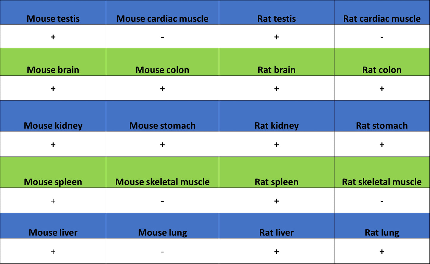
IHC shows positive staining in paraffin-embedded mouse testis. Anti-GFPT1 antibody was used at 1/500 dilution, followed by a HRP Polymer for Rabbit IgG (ready to use). Counterstained with hematoxylin. Heat mediated antigen retrieval with Tris/EDTA buffer pH9.0 was performed before commencing with IHC staining protocol.
IHC shows positive staining in paraffin-embedded rat testis. Anti-GFPT1 antibody was used at 1/500 dilution, followed by a HRP Polymer for Rabbit IgG (ready to use). Counterstained with hematoxylin. Heat mediated antigen retrieval with Tris/EDTA buffer pH9.0 was performed before commencing with IHC staining protocol.