WB result of GFP Recombinant Rabbit mAb
Lysate: Aequorea victoria GFP Protein 20ng
Primary antibody:
Lane 1: GFP Rabbit Recombinant mAb at 1/5000 dilution
Lane 2: GFP Rabbit Recombinant mAb at 1/20000 dilution
Lane 3: GFP Rabbit Recombinant mAb at 1/50000 dilution
Lane 4: GFP Rabbit Recombinant mAb at 1/100000 dilution
Secondary antibody: Goat Anti-Rabbit IgG, (H+L), HRP conjugated at 1/10000 dilution
Predicted MW: 28 kDa
Observed MW: 28 kDa
GFP Recombinant Rabbit mAb (S-296-169)
GFP Recombinant Rabbit mAb (S-296-169)
Price:
Regular price
$70 USD
Regular price
Sale price
$70 USD
Unit price
per
For shipping services or bulk orders, you may request a quotation.
Secure checkout with

View full details
Product Details
Product Details
Product Specification
| Host | Rabbit |
| Antigen | GFP |
| Immunogen | Synthetic Peptide |
| Accession | P42212 |
| Clone Number | S-296-169 |
| Antibody Type | Recombinant mAb |
| Isotype | IgG |
| Application | WB, ChIP |
| Reactivity | Species Independent |
| Cross Reactivity | This antibody is reactive against all variants of Aequorea victoria GFP such as CFP, ECFP, YFP, BFP, EGFP, Clover, EYFP and EVenus. |
| Purification | Protein A |
| Concentration | 0.5 mg/ml |
| Conjugation | Unconjugated |
| Physical Appearance | Liquid |
| Storage Buffer | PBS, 40% Glycerol, 0.05% BSA, 0.03% Proclin 300 |
| Stability & Storage | 12 months from date of receipt / reconstitution, -20 °C as supplied. |
Dilution
| application | dilution | species |
| WB | 1:50000-1:100000 | Species Independent |
Background
Green Fluorescent Protein (GFP) is a bioluminescent polypeptide consisting of 238 residues, originally isolated from the jellyfish Aequorea victoria. GFP converts the blue chemiluminescent light of aequorin into green fluorescent light, with an excitation peak at 395 nm and a minor peak at 475 nm, and an emission peak at 508 nm. The protein's structure includes a β-barrel with an α-helix running through its center, containing the fluorophore responsible for its fluorescence. GFP has become a crucial tool in research for monitoring gene expression, cellular localization, protein mobility, and interactions between various proteins. Mutant forms of GFP have been engineered to improve its photostability and to produce different colors, expanding its utility in various biological systems.
Picture
Picture
Western Blot
ChIP
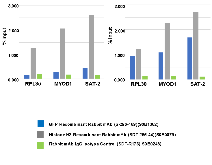
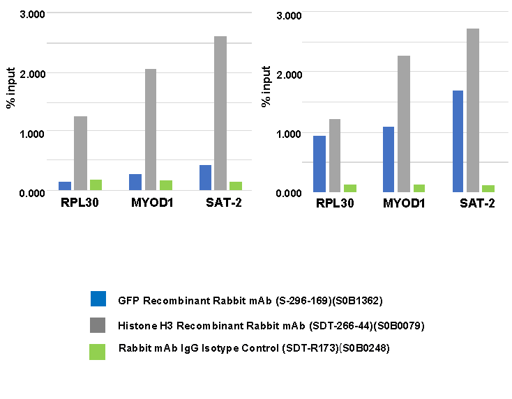
Chromatin immunoprecipitation (ChIP) was
performed on 293T cells were either untransfected
(left panel) or transfected with an GFP-tagged human
H3 construct (right panel) cross - linked with 1%
formaldehyde for 10 min, then chromatin was
fragmented by sonication.
Parallel reactions used GFP Recombinant Rabbit mAb
(S-296-169), Histone H3 Recombinant Rabbit mAb
(SDT-266-44) and Rabbit mAb IgG Isotype
Control (SDT-R173) at 1:50 for immunoprecipitation.
Post - immunoprecipitation, both samples were washed,
eluted, and cross - links reversed. Purified DNA was
analyzed by qPCR.
qPCR (%input: immunoprecipitated DNA/input DNA)
showed the enrichment of RPL30, MYOD1 and SAT-2 in
GFP Recombinant Rabbit mAb (S-296-169)-
immunoprecipitated sample.
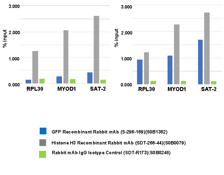
Chromatin immunoprecipitation (ChIP) was
performed on 293T cells were either untransfected
(left panel) or transfected with an GFP-tagged human
H3 construct (right panel) cross - linked with 1%
formaldehyde for 10 min, then chromatin was
fragmented by sonication.
Parallel reactions used GFP Recombinant Rabbit mAb
(S-296-169), Histone H3 Recombinant Rabbit mAb
(SDT-266-44) and Rabbit mAb IgG Isotype
Control (SDT-R173) at 1:50 for immunoprecipitation.
Post - immunoprecipitation, both samples were washed,
eluted, and cross - links reversed. Purified DNA was
analyzed by qPCR.
qPCR (%input: immunoprecipitated DNA/input DNA)
showed the enrichment of RPL30, MYOD1 and SAT-2 in
GFP Recombinant Rabbit mAb (S-296-169)-
immunoprecipitated sample.