WB result of GEF H1 Recombinant Rabbit mAb
Primary antibody: GEF H1 Recombinant Rabbit mAb at 1/1000 dilution
Lane 1: HeLa whole cell lysate 20 μg
Lane 2: U-87 MG whole cell lysate 20 μg
Lane 3: PC-3 whole cell lysate 20 μg
Secondary antibody: Goat Anti-Rabbit IgG (H+L), HRP conjugated at 1/10000 dilution
Predicted MW: 112 kDa
Observed MW: 112 kDa
GEF H1 Recombinant Rabbit mAb (S-6057)
GEF H1 Recombinant Rabbit mAb (S-6057)
Price:
Regular price
$100 USD
Regular price
Sale price
$100 USD
Unit price
per
For shipping services or bulk orders, you may request a quotation.
Secure checkout with

View full details
Product Details
Product Details
Product Specification
| Host | Rabbit |
| Antigen | GEF H1 |
| Synonyms | Rho guanine nucleotide exchange factor 2; Guanine nucleotide exchange factor H1 (GEF-H1); Microtubule-regulated Rho-GEF; Proliferating cell nucleolar antigen p40; KIAA0651; LFP40; ARHGEF2 |
| Location | Cytoplasm, Cytoskeleton |
| Accession | Q92974 |
| Clone Number | S-6057 |
| Antibody Type | Recombinant mAb |
| Isotype | IgG |
| Application | WB, ICC |
| Reactivity | Hu, Ms, Rt |
| Positive Sample | HeLa, U-87 MG, PC-3, RAW264.7, NIH/3T3, mouse brain, C6, rat brain |
| Purification | Protein A |
| Concentration | 0.5 mg/ml |
| Conjugation | Unconjugated |
| Physical Appearance | Liquid |
| Storage Buffer | PBS, 40% Glycerol, 0.05% BSA, 0.02% sodium azide |
| Stability & Storage | 12 months from date of receipt / reconstitution, -20 °C as supplied |
Dilution
| application | dilution | species |
| WB | 1:1000 | Hu, Ms, Rt |
| ICC | 1:500 | Hu |
Background
GEF-H1 (Guanine nucleotide exchange factor H1), encoded by the gene ARHGEF2, is a functionally critical Rho guanine nucleotide exchange factor (RhoGEF) whose core function is to catalyze the conversion of the small GTPase RhoA from an inactive GDP-bound state to an active GTP-bound state, thereby activating downstream signaling pathways that regulate actin cytoskeleton reorganization and a variety of cellular functions. Its activity is subject to complex regulation: on one hand, its protein structure contains typical domains such as DH (Dbl homology) and PH (pleckstrin homology), conferring catalytic activity and membrane localization capabilities; on the other hand, it can bind to microtubules, and upon microtubule depolymerization, GEF-H1 is released from microtubules and activated, thereby tightly coupling dynamic changes in the cytoskeleton to the switching of RhoA signaling pathways. In terms of physiological functions, GEF-H1 participates in regulating cell morphology, cell division, immune responses, and synaptic plasticity in neurons—for example, in neurons, it serves as a component of the AMPA receptor complex and negatively regulates dendritic spine development through RhoA signaling cascades. However, its functional dysregulation is also closely associated with various pathological conditions, such as hyperactivation of the GEF-H1/RhoA pathway observed in kidney diseases, and its involvement in regulating NF-κB activity and the production of inflammatory cytokines (such as IL-6 and TNF) also suggests its potential role in inflammatory diseases.
Picture
Picture
Western Blot
WB result of GEF H1 Recombinant Rabbit mAb
Primary antibody: GEF H1 Recombinant Rabbit mAb at 1/1000 dilution
Lane 1: Raw 264.7 whole cell lysate 20 μg
Lane 2: NIH/3T3 whole cell lysate 20 μg
Lane 3: mouse brain lysate 20 μg
Secondary antibody: Goat Anti-Rabbit IgG (H+L), HRP conjugated at 1/10000 dilution
Predicted MW: 112 kDa
Observed MW: 112 kDa
This blot was developed with high sensitivity substrate
WB result of GEF H1 Recombinant Rabbit mAb
Primary antibody: GEF H1 Recombinant Rabbit mAb at 1/1000 dilution
Lane 1: C6 whole cell lysate 20 μg
Lane 2: rat brain lysate 20 μg
Secondary antibody: Goat Anti-Rabbit IgG (H+L), HRP conjugated at 1/10000 dilution
Predicted MW: 112 kDa
Observed MW: 112 kDa
Immunocytochemistry
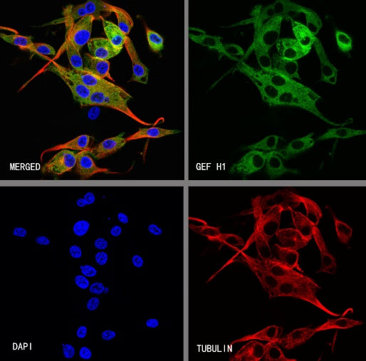
ICC shows positive staining in U-87 MG cells. Anti-GEF H1 antibody was used at 1/500 dilution (Green) and incubated overnight at 4°C. Goat polyclonal Antibody to Rabbit IgG - H&L (Alexa Fluor® 488) was used as secondary antibody at 1/1000 dilution. The cells were fixed with 4% PFA and permeabilized with 0.1% PBS-Triton X-100. Nuclei were counterstained with DAPI (Blue). Counterstain with tubulin (Red).