WB result of GATA1 Recombinant Rabbit mAb
Primary antibody: GATA1 Recombinant Rabbit mAb at 1/1000 dilution
Lane 1: HeLa whole cell lysate 20 µg
Lane 2: K-562 whole cell lysate 20 µg
Negative control: HeLa whole cell lysate
Secondary antibody: Goat Anti-Rabbit IgG, (H+L) HRP conjugated at 1/10000 dilution
Predicted MW: 43 kDa
Observed MW: 43 kDa
GATA1 Recombinant Rabbit mAb (S-5401)
GATA1 Recombinant Rabbit mAb (S-5401)
Price:
Regular price
$100 USD
Regular price
Sale price
$100 USD
Unit price
per
For shipping services or bulk orders, you may request a quotation.
Secure checkout with

View full details
Product Details
Product Details
Product Specification
| Host | Rabbit |
| Antigen | GATA1 |
| Synonyms | Erythroid transcription factor; Eryf1; GATA-binding factor 1 (GATA-1; GF-1); NF-E1 DNA-binding protein; GF1; GATA1 |
| Location | Nucleus |
| Accession | P15976 |
| Clone Number | S-5401 |
| Antibody Type | Recombinant mAb |
| Isotype | IgG |
| Application | WB, IHC-P, ICC |
| Reactivity | Hu, Ms, Rt |
| Positive Sample | K-562 |
| Purification | Protein A |
| Concentration | 0.5 mg/ml |
| Conjugation | Unconjugated |
| Physical Appearance | Liquid |
| Storage Buffer | PBS, 40% Glycerol, 0.05% BSA, 0.02% sodium azide |
| Stability & Storage | 12 months from date of receipt / reconstitution, -20 °C as supplied |
Dilution
| application | dilution | species |
| WB | 1:1000 | Hu, Ms, Rt |
| IHC-P | 1:500 | Hu, Ms, Rt |
| ICC | 1:500 | Hu |
Background
GATA1 is a zinc finger transcription factor that serves as the master regulator of erythroid and megakaryocytic differentiation, playing an indispensable role in hematopoiesis by binding to the consensus DNA sequence (A/T)GATA(A/G) to activate genes essential for red blood cell maturation, such as those encoding globins and heme biosynthesis enzymes, while simultaneously repressing proliferation-associated genes to facilitate terminal differentiation. Its activity is tightly controlled through post-translational modifications and critical protein-protein interactions with cofactors like FOG-1 (ZNF423), which modulates its transcriptional output, and mutations in the GATA1 gene are clinically significant as they cause X-linked dyserythropoietic anemia, thrombocytopenia, and are strongly associated with transient abnormal myelopoiesis and acute megakaryoblastic leukemia in individuals with Down syndrome, underscoring its vital function in normal blood development and its pathological consequences when dysregulated.
Picture
Picture
Western Blot
Immunohistochemistry
IHC shows positive staining in paraffin-embedded human colon cancer. Anti-GATA1 antibody was used at 1/500 dilution, followed by a HRP Polymer for Rabbit IgG (ready to use). Counterstained with hematoxylin. Heat mediated antigen retrieval with Tris/EDTA buffer pH9.0 was performed before commencing with IHC staining protocol.
IHC shows positive staining in paraffin-embedded human lung cancer. Anti-GATA1 antibody was used at 1/500 dilution, followed by a HRP Polymer for Rabbit IgG (ready to use). Counterstained with hematoxylin. Heat mediated antigen retrieval with Tris/EDTA buffer pH9.0 was performed before commencing with IHC staining protocol.
IHC shows positive staining in paraffin-embedded mouse bone marrow. Anti-GATA1 antibody was used at 1/500 dilution, followed by a HRP Polymer for Rabbit IgG (ready to use). Counterstained with hematoxylin. Heat mediated antigen retrieval with Tris/EDTA buffer pH9.0 was performed before commencing with IHC staining protocol.
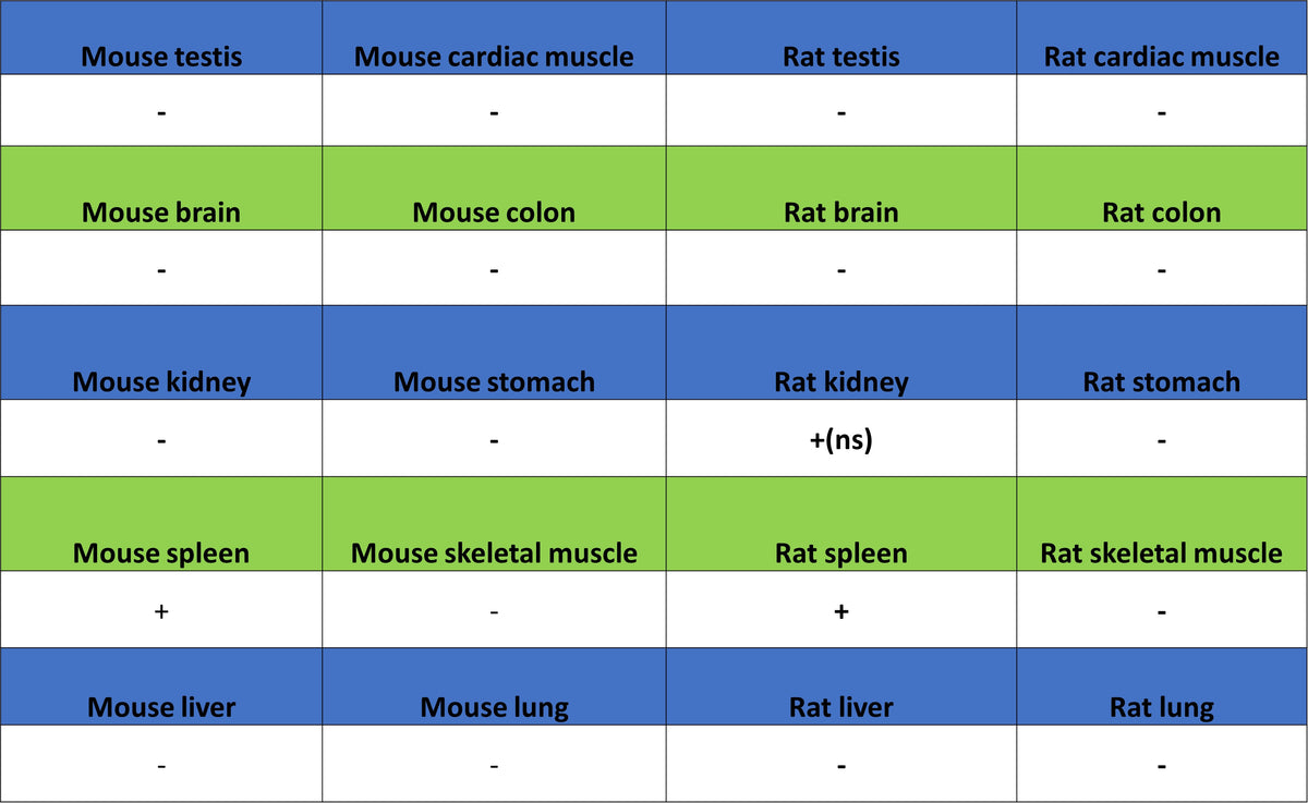
IHC shows positive staining in paraffin-embedded rat spleen. Anti-GATA1 antibody was used at 1/500 dilution, followed by a HRP Polymer for Rabbit IgG (ready to use). Counterstained with hematoxylin. Heat mediated antigen retrieval with Tris/EDTA buffer pH9.0 was performed before commencing with IHC staining protocol.
Immunocytochemistry
ICC shows positive staining in K562 cells (top panel) and negative staining in HeLa cells (below panel). Anti-GATA1 antibody was used at 1/500 dilution (Green) and incubated overnight at 4°C. Goat polyclonal Antibody to Rabbit IgG - H&L (Alexa Fluor® 488) was used as secondary antibody at 1/1000 dilution. The cells were fixed with 4% PFA and permeabilized with 0.1% PBS-Triton X-100. Nuclei were counterstained with DAPI (Blue). Counterstain with tubulin (Red).