GAB1 Recombinant Rabbit mAb (S-3907-33)
GAB1 Recombinant Rabbit mAb (S-3907-33)
Price:
Regular price
$100 USD
Regular price
Sale price
$100 USD
Unit price
per
For shipping services or bulk orders, you may request a quotation.
Secure checkout with

View full details
Product Details
Product Details
Product Specification
| Host | Rabbit |
| Antigen | GAB1 |
| Synonyms | GRB2-associated-binding protein 1; GRB2-associated binder 1; Growth factor receptor bound protein 2-associated protein 1 |
| Immunogen | Synthetic Peptide |
| Location | Cytoplasm |
| Accession | Q13480 |
| Clone Number | S-3907-33 |
| Antibody Type | Recombinant mAb |
| Isotype | IgG |
| Application | WB, IHC-P |
| Reactivity | Hu, Ms, Mk |
| Positive Sample | Ramos, HepG2, NIH/3T3, 3T3/L1, mouse brain, COS7 |
| Predicted Reactivity | Bv, Hm |
| Purification | Protein A |
| Concentration | 0.5 mg/ml |
| Conjugation | Unconjugated |
| Physical Appearance | Liquid |
| Storage Buffer | PBS, 40% Glycerol, 0.05% BSA, 0.02% sodium azide |
| Stability & Storage | 12 months from date of receipt / reconstitution, -20 °C as supplied |
Dilution
| application | dilution | species |
| WB | 1:1000 | Hu, Ms, Mk |
| IHC-P | 1:250 | Hu |
Background
GAB1 is a core member of the Gab family of adaptor proteins, consisting of 694 amino acids with a molecular weight of approximately 77 kDa. As a multifunctional adapter protein, GAB1 lacks catalytic activity but serves as a critical bridging molecule in intracellular signal transduction through its N-terminal pleckstrin homology (PH) domain, proline-rich regions, and multiple tyrosine residues that can be phosphorylated. It can be activated by various receptor tyrosine kinases such as the epidermal growth factor receptor (EGFR) and the hepatocyte growth factor receptor (c-Met), and by binding to effector molecules including GRB2, the p85 subunit of PI3K, and the phosphatase SHP2, it mediates the activation of downstream signaling pathways such as PI3K-AKT and MAPK, thereby broadly participating in the regulation of cell growth, proliferation, migration, and apoptosis. Notably, GAB1 can directly bind to the c-Met receptor through its unique Met-binding domain (MBD), a feature that renders it an irreplaceable role in mediating hepatocyte growth factor (HGF)-induced cell branching morphogenesis and angiogenesis. Furthermore, functional aberrations or altered expression of GAB1 have been implicated in various diseases, including tumor development and progression, inflammatory responses, and hereditary non-syndromic deafness.
Picture
Picture
Western Blot
WB result of GAB1 Recombinant Rabbit mAb
Primary antibody: GAB1 Recombinant Rabbit mAb at 1/1000 dilution
Lane 1: Ramos whole cell lysate 20 µg
Lane 2: HepG2 whole cell lysate 20 µg
Secondary antibody: Goat Anti-Rabbit IgG (H+L), HRP conjugated at 1/10000 dilution
Predicted MW: 77 kDa
Observed MW: 100 kDa
This blot was developed with high sensitivity substrate
WB result of GAB1 Recombinant Rabbit mAb
Primary antibody: GAB1 Recombinant Rabbit mAb at 1/1000 dilution
Lane 1: NIH/3T3 whole cell lysate 20 µg
Lane 2: 3T3/L1 whole cell lysate 20 µg
Lane 3: mouse brain lysate 20 µg
Secondary antibody: Goat Anti-Rabbit IgG (H+L), HRP conjugated at 1/10000 dilution
Predicted MW: 77 kDa
Observed MW: 100 kDa
WB result of GAB1 Recombinant Rabbit mAb
Primary antibody: GAB1 Recombinant Rabbit mAb at 1/1000 dilution
Lane 1: COS7 whole cell lysate 20 µg
Secondary antibody: Goat Anti-Rabbit IgG (H+L), HRP conjugated at 1/10000 dilution
Predicted MW: 77 kDa
Observed MW: 100 kDa
This blot was developed with high sensitivity substrate
Immunohistochemistry
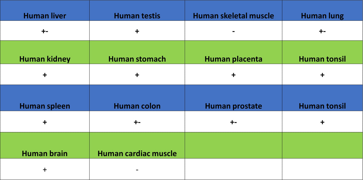
IHC shows positive staining in paraffin-embedded human kidney. Anti-GAB1 antibody was used at 1/250 dilution, followed by a HRP Polymer for Rabbit IgG (ready to use). Counterstained with hematoxylin. Heat mediated antigen retrieval with Tris/EDTA buffer pH9.0 was performed before commencing with IHC staining protocol.
IHC shows positive staining in paraffin-embedded human placenta. Anti-GAB1 antibody was used at 1/250 dilution, followed by a HRP Polymer for Rabbit IgG (ready to use). Counterstained with hematoxylin. Heat mediated antigen retrieval with Tris/EDTA buffer pH9.0 was performed before commencing with IHC staining protocol.
IHC shows positive staining in paraffin-embedded human breast cancer. Anti-GAB1 antibody was used at 1/250 dilution, followed by a HRP Polymer for Rabbit IgG (ready to use). Counterstained with hematoxylin. Heat mediated antigen retrieval with Tris/EDTA buffer pH9.0 was performed before commencing with IHC staining protocol.
IHC shows positive staining in paraffin-embedded human lung cancer. Anti-GAB1 antibody was used at 1/250 dilution, followed by a HRP Polymer for Rabbit IgG (ready to use). Counterstained with hematoxylin. Heat mediated antigen retrieval with Tris/EDTA buffer pH9.0 was performed before commencing with IHC staining protocol.