WB result of FOLR1 Recombinant Rabbit mAb
Primary antibody: FOLR1 Recombinant Rabbit mAb at 1/1000 dilution
Lane 1: K562 whole cell lysate 20 µg
Lane 2: JAR whole cell lysate 20 µg
Lane 3: HeLa whole cell lysate 20 µg
Lane 4: SK-OV-3 whole cell lysate 20 µg
Lane 5: OVCAR-3 whole cell lysate 20 µg
Negative control: K562 whole cell lysate
Secondary antibody: Goat Anti- rabbit IgG, (H+L), HRP conjugated at 1/10000 dilution
Predicted MW: 30 kDa
Observed MW: 36 kDa
FOLR1 Recombinant Rabbit mAb (SDT-3635-124)
FOLR1 Recombinant Rabbit mAb (SDT-3635-124)
Price:
Regular price
$100 USD
Regular price
Sale price
$100 USD
Unit price
per
For shipping services or bulk orders, you may request a quotation.
Secure checkout with

View full details
Product Details
Product Details
Product Specification
| Host | Rabbit |
| Antigen | FOLR1 |
| Synonyms | Folate receptor alpha; FR-alpha; Adult folate-binding protein (FBP); Folate receptor 1; Folate receptor; adult; KB cells FBP; Ovarian tumor-associated antigen MOv18; FOLR |
| Immunogen | Recombinant Protein |
| Location | Secreted, Endosome, Cell membrane |
| Accession | P15328 |
| Clone Number | SDT-3635-124 |
| Antibody Type | Recombinant mAb |
| Isotype | IgG |
| Application | WB, IHC-P |
| Reactivity | Hu, Ms, Rt |
| Positive Sample | JAR, HeLa, SK-OV-3, OVCAR-3 |
| Purification | Protein A |
| Concentration | 0.5 mg/ml |
| Conjugation | Unconjugated |
| Physical Appearance | Liquid |
| Storage Buffer | PBS, 40% Glycerol, 0.05% BSA, 0.02% sodium azide |
| Stability & Storage | 12 months from date of receipt / reconstitution, -20 °C as supplied |
Dilution
| application | dilution | species |
| WB | 1:1000 | Hu, Ms |
| IHC-P | 1:500 | Hu, Ms, Rt |
Background
Human folate receptor alpha (FOLR1) is a glycoprotein that plays a crucial role in the transport of folate, an essential vitamin, into cells. This receptor is highly expressed in certain tissues and is particularly notable for its involvement in various physiological processes including embryonic development and cellular proliferation. Its overexpression has been observed in numerous cancers, such as ovarian, lung, and breast cancer, making it a potential target for cancer diagnosis and targeted therapy. By binding to folate or folate analogs, FOLR1 facilitates the internalization of these molecules via endocytosis, thereby providing cells with the necessary folate for metabolic functions like DNA synthesis and repair.
Picture
Picture
Western Blot
WB result of FOLR1 Recombinant Rabbit mAb
Primary antibody: FOLR1 Recombinant Rabbit mAb at 1/1000 dilution
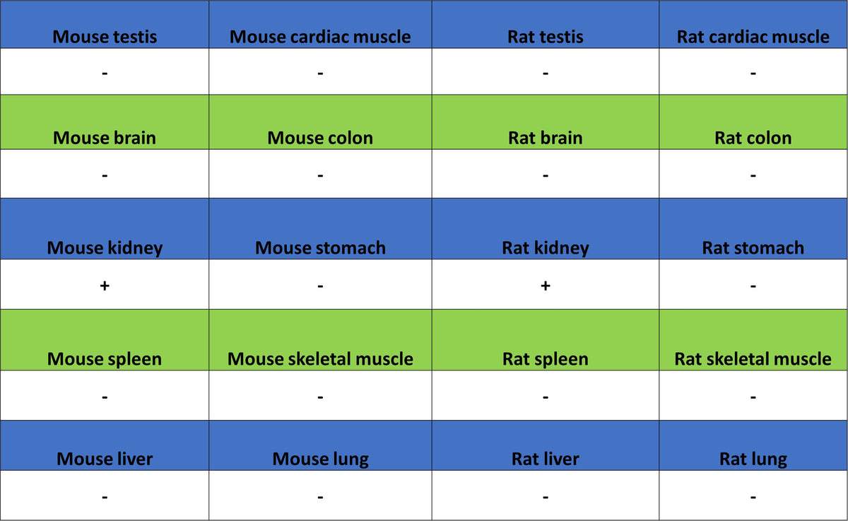
Lane 1: mouse kidney lysate 20 µg
Secondary antibody: Goat Anti- rabbit IgG, (H+L), HRP conjugated at 1/10000 dilution
Predicted MW: 30 kDa
Observed MW: 35 kDa
WB result of FOLR1 Recombinant Rabbit mAb
Primary antibody: FOLR1 Recombinant Rabbit mAb at 1/1000 dilution
Lane 1: rat kidney lysate 20 µg
Secondary antibody: Goat Anti- rabbit IgG, (H+L), HRP conjugated at 1/10000 dilution
Predicted MW: 30 kDa
Observed MW: 32 kDa
Immunohistochemistry
IHC shows positive staining in paraffin-embedded human kidney. Anti-FOLR1 antibody was used at 1/500 dilution, followed by a HRP Polymer for Rabbit IgG (ready to use). Counterstained with hematoxylin. Heat mediated antigen retrieval with Tris/EDTA buffer pH9.0 was performed before commencing with IHC staining protocol.
IHC shows positive staining in paraffin-embedded human ovarian cancer (case 1). Anti-FOLR1 antibody was used at 1/500 dilution, followed by a HRP Polymer for Rabbit IgG (ready to use). Counterstained with hematoxylin. Heat mediated antigen retrieval with Tris/EDTA buffer pH9.0 was performed before commencing with IHC staining protocol.
IHC shows positive staining in paraffin-embedded human ovarian cancer (case 2). Anti-FOLR1 antibody was used at 1/500 dilution, followed by a HRP Polymer for Rabbit IgG (ready to use). Counterstained with hematoxylin. Heat mediated antigen retrieval with Tris/EDTA buffer pH9.0 was performed before commencing with IHC staining protocol.
IHC shows positive staining in paraffin-embedded mouse kidney. Anti-FOLR1 antibody was used at 1/500 dilution, followed by a HRP Polymer for Rabbit IgG (ready to use). Counterstained with hematoxylin. Heat mediated antigen retrieval with Tris/EDTA buffer pH9.0 was performed before commencing with IHC staining protocol.
IHC shows positive staining in paraffin-embedded rat kidney. Anti-FOLR1 antibody was used at 1/500 dilution, followed by a HRP Polymer for Rabbit IgG (ready to use). Counterstained with hematoxylin. Heat mediated antigen retrieval with Tris/EDTA buffer pH9.0 was performed before commencing with IHC staining protocol.