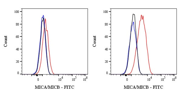
Flow cytometric analysis of Human MICA/MICB expression on HeLa cells. Cells from the HeLa (Human monocytic leukemia monocyte, right) or THP-1 (Human prostate adenocarcinoma epithelial cell, left) were stained with either FITC Mouse IgG2a, κ Isotype Control (black line histogram) or SDT FITC Mouse Anti-Human MICA/MICB Antibody (red line histogram) at 5 μl/test followed by Sav-iFluor 488, cells without incubation with primary antibody and secondary antibody (blue line histogram) were used as unlabeled control. Flow cytometry and data analysis were performed using Agilent NovoCyte Quanteon and FlowJo™ software.
FITC Mouse Anti-Human MICA/MICB Antibody (6D4)
FITC Mouse Anti-Human MICA/MICB Antibody (6D4)
Price:
Regular price
$195 USD
Regular price
Sale price
$195 USD
Unit price
per
For shipping services or bulk orders, you may request a quotation.
Secure checkout with

View full details
Product Details
Product Details
Product Specification
| Host | Mouse |
| Antigen | MICA/MICB |
| Location | Cell membrane |
| Accession | Q29983、 Q29980 |
| Clone Number | 6D4 |
| Antibody Type | Mouse mAb |
| Isotype Control | S0B5453 |
| Application | ICFCM |
| Reactivity | Hu |
| Positive Sample | HeLa |
| Purification | Protein A |
| Concentration | 0.2 mg/ml |
| Conjugation | FITC |
| Physical Appearance | Liquid |
| Storage Buffer | PBS, 1% BSA, 0.09% sodium azide |
| Stability & Storage | 12 months from date of receipt / reconstitution, 2 to 8 °C as supplied |
Dilution
| application | dilution | species |
| ICFCM | 5μl per million cells in 100μl volume | Hu |
Background
MICA (MHC class I polypeptide-related sequence A) and MICB (MHC class I polypeptide-related sequence B) are stress-inducible proteins that belong to the MHC class I family but do not bind to β2-microglobulin or present peptides. They are encoded by genes located within the HLA class I region of chromosome 6. MICA and MICB are expressed on the cell membrane in response to cellular stress or cancer transformation. These proteins act as ligands for the NKG2D receptor, which is expressed on most natural killer (NK) cells, CD8 αβ T cells, and γδ T cells. The binding of MICA/B to NKG2D can activate these immune cells, leading to cytotoxic responses against stressed or transformed cells. MICA and MICB are highly polymorphic, with over 106 alleles reported for MICA and 25 for MICB. This polymorphism may affect their affinity for NKG2D and, consequently, the activation thresholds of NK and T cells.
Picture
Picture
FC