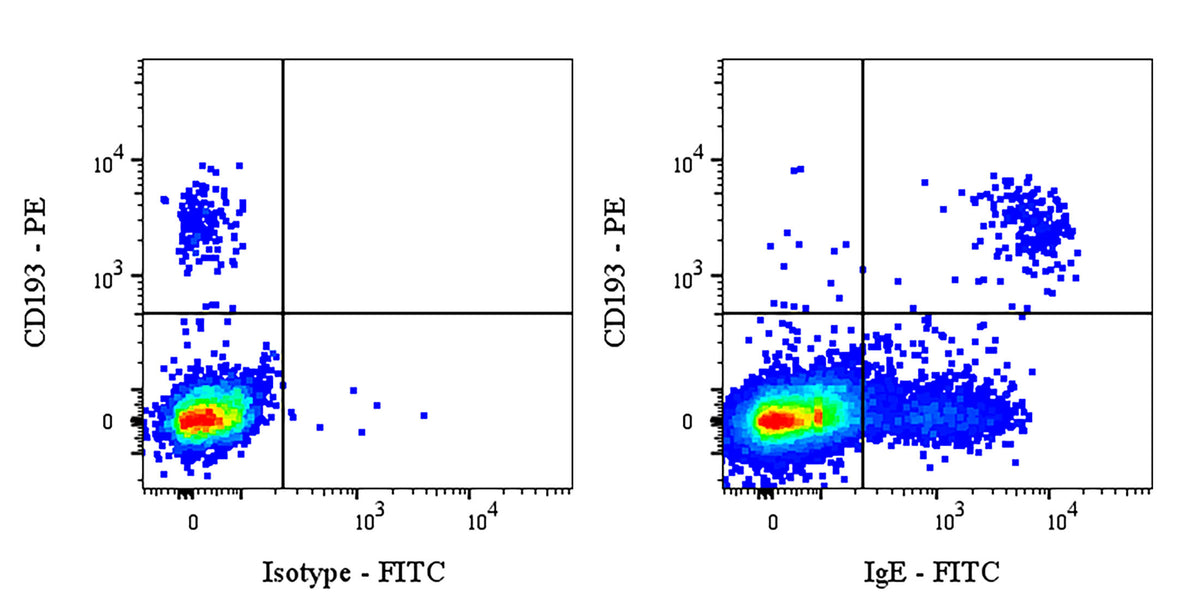
Flow cytometric analysis of Human IgE expression on Human PBMCs. Human PBMCs (peripheral blood mononuclear cells) were stained with PE Mouse Anti-Human CD193 Antibody and either FITC Mouse IgG1, κ Isotype Control (left panel) or SDT FITC Mouse Anti-Human IgE Antibody (right panel) at 5 μl/test. Flow cytometry and data analysis were performed using BD FACSymphony™ A1 and FlowJo™ software.
FITC Mouse Anti-Human IgE Antibody (S-5486)
FITC Mouse Anti-Human IgE Antibody (S-5486)
Price:
Regular price
$200 USD
Regular price
Sale price
$200 USD
Unit price
per
For shipping services or bulk orders, you may request a quotation.
Secure checkout with

View full details
Product Details
Product Details
Product Specification
| Host | Mouse |
| Antigen | IgE |
| Synonyms | Immunoglobulin epsilon heavy chain |
| Location | Secreted, Cell membrane |
| Accession | P0DOX4 |
| Clone Number | S-5486 |
| Antibody Type | Mouse mAb |
| Isotype | IgG1,k |
| Isotype Control | S0B1529 |
| Application | FCM |
| Reactivity | Hu |
| Positive Sample | Human PBMC |
| Purification | Protein G |
| Concentration | 0.2 mg/ml |
| Conjugation | FITC |
| Physical Appearance | Liquid |
| Storage Buffer | PBS, 1% BSA, 0.09% sodium azide |
| Stability & Storage | 12 months from date of receipt / reconstitution, 2 to 8 °C as supplied |
Dilution
| application | dilution | species |
| FCM | 5μl per million cells in 100μl volume | Hu |
Background
Immunoglobulin E (IgE) is the antibody class with the lowest serum concentration among the five immunoglobulin isotypes (approximately 0.05 μg/mL), yet it is highly conserved throughout evolution. Its basic structure is a tetrapeptide chain monomer composed of two identical heavy chains (ε chains) and two identical light chains linked by disulfide bonds. The heavy chain contains four constant region domains (Cε1–Cε4), among which the Cε3 domain serves as the core site for binding to the high-affinity FcεRI receptor, while the flexible hinge region between Cε2 and Cε3 confers unique conformational flexibility to the molecule. IgE is primarily produced by plasma cells in mucosal lymphoid tissues, and its unique biological functions are manifested in two major aspects. First, it acts as the "mastermind" of allergic reactions: upon initial exposure to an allergen, it induces B-cell class switching to produce specific IgE, which then sensitizes mast cells and basophils by binding to the high-affinity FcεRI receptor on their surfaces via its Fc portion. When the same allergen re-enters the body, it cross-links adjacent IgE molecules on the cell surface, triggering intracellular signaling cascades that lead to the release of preformed inflammatory mediators such as histamine and leukotrienes, as well as the de novo synthesis of cytokines (e.g., IL-4, IL-13), thereby eliciting a range of hypersensitivity reactions from local wheal-and-flare responses and pruritus to systemic anaphylactic shock. Second, IgE serves as a "guardian" in anti-parasitic immunity: through FcεRI-mediated antibody-dependent cell-mediated cytotoxicity (ADCC), IgE cooperates with eosinophils to release effector molecules such as major basic protein and peroxidases, effectively killing large parasites such as helminths. Furthermore, parasite-specific IgE can also promote antigen presentation via the low-affinity receptor FcεRII (CD23), amplifying Th2-type immune responses. At the clinical level, elevated total serum IgE levels often indicate an atopic predisposition (e.g., asthma, allergic rhinitis, atopic dermatitis) or can serve as an auxiliary diagnostic marker for parasitic infections. Monoclonal antibody drugs targeting IgE, such as Omalizumab, which binds to the Cε3 region of free IgE to block its interaction with FcεRI, have become effective targeted therapies for moderate-to-severe allergic asthma and chronic spontaneous urticaria. It is worth noting that dysregulated IgE production is also associated with certain autoimmune diseases and some types of tumors (e.g., hypereosinophilic syndrome); however, its precise function under normal physiological conditions and its evolutionary conservation remain frontier questions actively explored in the field of immunology.
Picture
Picture
FC