Ferritin Heavy Chain Recombinant Rabbit mAb (S-5419)
Ferritin Heavy Chain Recombinant Rabbit mAb (S-5419)
Price:
Regular price
$100 USD
Regular price
Sale price
$100 USD
Unit price
per
For shipping services or bulk orders, you may request a quotation.
Secure checkout with

View full details
Product Details
Product Details
Product Specification
| Host | Rabbit |
| Antigen | Ferritin Heavy Chain |
| Synonyms | Ferritin H subunit; HftCleaved into 1 chainFerritin heavy chain; N-terminally processed; Fth; Fth1 |
| Location | Cytoplasm, Lysosome |
| Accession | P09528 |
| Clone Number | S-5419 |
| Antibody Type | Recombinant mAb |
| Isotype | IgG |
| Application | WB, IHC-P |
| Reactivity | Ms, Rt |
| Positive Sample | RAW264.7, NIH/3T3, mouse liver, mouse brain, mouse heart, mouse spleen, PC-12, C6, rat live, rat brain, rat kidney, rat spleen |
| Purification | Protein A |
| Concentration | 0.5 mg/ml |
| Conjugation | Unconjugated |
| Physical Appearance | Liquid |
| Storage Buffer | PBS, 40% Glycerol, 0.05% BSA, 0.02% sodium azide |
| Stability & Storage | 12 months from date of receipt / reconstitution, -20 °C as supplied |
Dilution
| application | dilution | species |
| WB | 1:1000 | Ms, Rt |
| IHC-P | 1:1000 | Ms, Rt |
Background
Ferritin Heavy Chain (FTH1) is the catalytic core subunit of the ferritin complex. Through its unique ferroxidase activity, it rapidly oxidizes toxic ferrous iron (Fe²⁺) to non-toxic ferric iron (Fe³⁺) and safely stores it within a spherical protein shell self-assembled from 24 subunits, thereby playing a critical role in maintaining cellular iron homeostasis and preventing oxidative damage. This sophisticated oxidative storage process not only protects cells from oxidative stress and ferroptosis triggered by free iron, but recent studies have also revealed its deep involvement in immune regulation and tissue repair—for example, in regulating macrophage function during kidney injury. Conversely, its dysfunction is closely linked to hereditary hemochromatosis and brain iron accumulation in neurodegenerative diseases.
Picture
Picture
Western Blot
WB result of Ferritin Heavy Chain Recombinant Rabbit mAb
Primary antibody: Ferritin Heavy Chain Recombinant Rabbit mAb at 1/1000 dilution
Lane 1: Raw 264.7 whole cell lysate 20 µg
Lane 2: NIH/3T3 whole cell lysate 20 µg
Lane 3: mouse liver lysate 20 µg
Lane 4: mouse brain lysate 20 µg
Lane 5: mouse heart lysate 20 µg
Lane 6: mouse spleen lysate 20 µg
Secondary antibody: Goat Anti-Rabbit IgG, (H+L), HRP conjugated at 1/10000 dilution
Predicted MW: 21 kDa
Observed MW: 22 kDa
WB result of Ferritin Heavy Chain Recombinant Rabbit mAb
Primary antibody: Ferritin Heavy Chain Recombinant Rabbit mAb at 1/1000 dilution
Lane 1: PC-12 whole cell lysate 20 µg
Lane 2: C6 whole cell lysate 20 µg
Lane 3: rat liver lysate 20 µg
Lane 4: rat brain lysate 20 µg
Lane 5: rat kidney lysate 20 µg
Lane 6: rat spleen lysate 20 µg
Secondary antibody: Goat Anti-Rabbit IgG, (H+L), HRP conjugated at 1/10000 dilution
Predicted MW: 21 kDa
Observed MW: 22 kDa
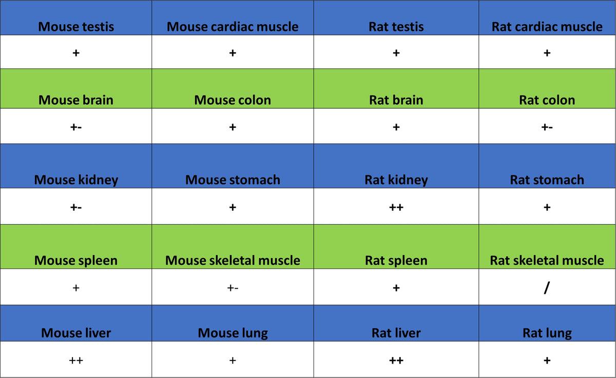
Immunohistochemistry
IHC shows positive staining in paraffin-embedded mouse liver. Anti-Ferritin heavy chain antibody was used at 1/1000 dilution, followed by a HRP Polymer for Rabbit IgG (ready to use). Counterstained with hematoxylin. Heat mediated antigen retrieval with Tris/EDTA buffer pH9.0 was performed before commencing with IHC staining protocol.
IHC shows positive staining in paraffin-embedded rat kidney. Anti-Ferritin heavy chain antibody was used at 1/1000 dilution, followed by a HRP Polymer for Rabbit IgG (ready to use). Counterstained with hematoxylin. Heat mediated antigen retrieval with Tris/EDTA buffer pH9.0 was performed before commencing with IHC staining protocol.
IHC shows positive staining in paraffin-embedded rat liver. Anti-Ferritin heavy chain antibody was used at 1/1000 dilution, followed by a HRP Polymer for Rabbit IgG (ready to use). Counterstained with hematoxylin. Heat mediated antigen retrieval with Tris/EDTA buffer pH9.0 was performed before commencing with IHC staining protocol.