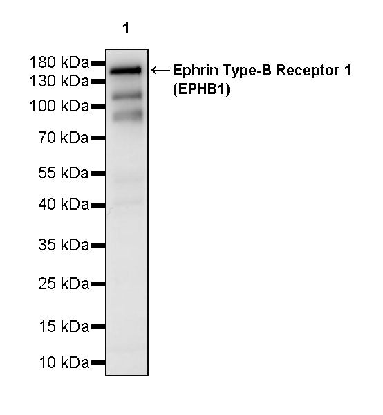
WB result of Ephrin Type-B Receptor 1 (EPHB1) Recombinant Rabbit mAb
Primary antibody: Ephrin Type-B Receptor 1 (EPHB1) Recombinant Rabbit mAb at 1/1000 dilution
Lane 1: SH-SY5Y whole cell lysate 20 µg
Secondary antibody: Goat Anti- rabbit IgG, (H+L), HRP conjugated at 1/10000 dilution
Predicted MW: 110 kDa
Observed MW: 150 kDa
This blot was developed with high sensitivity substrate
Ephrin Type-B Receptor 1 (EPHB1) Recombinant Rabbit mAb (S-4151-65)
Ephrin Type-B Receptor 1 (EPHB1) Recombinant Rabbit mAb (S-4151-65)
Price:
Regular price
$100 USD
Regular price
Sale price
$100 USD
Unit price
per
For shipping services or bulk orders, you may request a quotation.
Secure checkout with

View full details
Product Details
Product Details
Product Specification
| Host | Rabbit |
| Antigen | Ephrin Type-B Receptor 1 (EPHB1) |
| Synonyms | ELK; EPH tyrosine kinase 2; EPH-like kinase 6 (EK6; hEK6); Neuronally-expressed EPH-related tyrosine kinase (NET); Tyrosine-protein kinase receptor EPH-2; EPHT2; HEK6; NET |
| Immunogen | Synthetic Peptide |
| Location | Cell membrane, Endosome |
| Accession | P54762 |
| Clone Number | S-4151-65 |
| Antibody Type | Recombinant mAb |
| Isotype | IgG |
| Application | WB |
| Reactivity | Hu, Ms, Rt |
| Positive Sample | SH-SY5Y, mouse brain, rat brain |
| Purification | Protein A |
| Concentration | 0.5 mg/ml |
| Conjugation | Unconjugated |
| Physical Appearance | Liquid |
| Storage Buffer | PBS, 40% Glycerol, 0.05% BSA, 0.02% sodium azide |
| Stability & Storage | 12 months from date of receipt / reconstitution, -20 °C as supplied |
Dilution
| application | dilution | species |
| WB | 1:1000 | Hu, Ms, Rt |
Background
Ephrin Type-B Receptor 1 (EphB1) is an important member of the Eph family of receptor tyrosine kinases, primarily mediating cell-cell bidirectional signaling through interactions with membrane-anchored ephrin-B ligands, and playing critical regulatory roles in neurodevelopment, angiogenesis, and tumorigenesis. The canonical structure of this protein consists of a ligand-binding extracellular globular domain, a cysteine-rich region, two fibronectin type III repeats, a single transmembrane domain, and an intracellular tyrosine kinase domain, with the cytoplasmic region also containing multiple phosphorylatable tyrosine residues and a PDZ domain-binding motif. As a core component of axon guidance molecules, EphB1 mediates "forward signaling" through its kinase activity to regulate cytoskeletal rearrangement, while also receiving "reverse signals" carried by ephrin-B ligands that affect the adhesion and migration of ligand-expressing cells. In nervous system development, EphB1 participates in establishing retinotectal projections and corpus callosum connections by controlling growth cone collapse and axon pathfinding. Furthermore, accumulating studies have shown that EphB1 is aberrantly expressed in various solid tumors, influencing malignant progression and patient prognosis by promoting tumor angiogenesis, enhancing tumor cell invasiveness, and interacting with the immune microenvironment, thereby positioning it as a potential anti-cancer therapeutic target.
Picture
Picture
Western Blot
WB result of Ephrin Type-B Receptor 1 (EPHB1) Recombinant Rabbit mAb
Primary antibody: Ephrin Type-B Receptor 1 (EPHB1) Recombinant Rabbit mAb at 1/1000 dilution
Lane 1: mouse brain lysate 20 µg
Secondary antibody: Goat Anti- rabbit IgG, (H+L), HRP conjugated at 1/10000 dilution
Predicted MW: 110 kDa
Observed MW: 150 kDa
This blot was developed with high sensitivity substrate
WB result of Ephrin Type-B Receptor 1 (EPHB1) Recombinant Rabbit mAb
Primary antibody: Ephrin Type-B Receptor 1 (EPHB1) Recombinant Rabbit mAb at 1/1000 dilution
Lane 1: rat brain lysate 20 µg
Secondary antibody: Goat Anti- rabbit IgG, (H+L), HRP conjugated at 1/10000 dilution
Predicted MW: 110 kDa
Observed MW: 150 kDa
This blot was developed with high sensitivity substrate