WB result of EPCR/PROCR(CD201) Rabbit Polyclonal Antibody
Primary antibody: EPCR/PROCR(CD201) Rabbit Polyclonal Antibody at 1/1000 dilution
Lane 1: Jurkat whole cell lysate 20 μg
Lane 2: HUVEC whole cell lysate 20 μg
Negative control: Jurkat whole cell lysate
Secondary antibody: Goat Anti-Rabbit IgG (H+L), HRP conjugated at 1/10000 dilution
Predicted MW: 26 kDa
Observed MW: 35-40 kDa
EPCR/PROCR (CD201) Rabbit Polyclonal Antibody
EPCR/PROCR (CD201) Rabbit Polyclonal Antibody
Price:
Regular price
$100 USD
Regular price
Sale price
$100 USD
Unit price
per
For shipping services or bulk orders, you may request a quotation.
Secure checkout with

View full details
Product Details
Product Details
Product Specification
| Host | Rabbit |
| Antigen | EPCR/PROCR(CD201) |
| Immunogen | Recombinant Protein |
| Location | Membrane |
| Accession | Q9UNN8 |
| Antibody Type | Polyclonal antibody |
| Isotype | IgG |
| Application | WB, IHC-P, IF |
| Reactivity | Hu |
| Positive Sample | HUVEC |
| Purification | Immunogen Affinity |
| Concentration | 2 mg/ml |
| Conjugation | Unconjugated |
| Physical Appearance | Liquid |
| Storage Buffer | PBS, 40% Glycerol, 0.05% BSA, 0.02% sodium azide |
| Stability & Storage | 12 months from date of receipt / reconstitution, -20 °C as supplied |
Dilution
| application | dilution | species |
| WB | 1:1000-1:2000 | Hu |
| IHC-P | 1:500 | Hu |
| IF | 1:2000 | Hu |
Background
Endothelial protein C receptor (EPCR) is a type I transmembrane glycoprotein that shares sequence and three-dimensional structural homology with the MHC class I/CD1 family, but its antigen-binding groove binds phospholipids (typically phosphatidylcholine) rather than antigenic peptides. EPCR is primarily expressed on the surface of large vessel endothelial cells and has also been detected on hematopoietic stem cells, monocytes, smooth muscle cells, and various progenitor cells. Its classical function is to bind protein C and present it to the thrombin-thrombomodulin complex, increasing the efficiency of protein C activation by approximately 5- to 20-fold and thereby exerting anticoagulant effects. In addition, EPCR initiates cytoprotective signaling, including anti-inflammatory, anti-apoptotic, and endothelial barrier-protective effects, by mediating the cleavage of protease-activated receptor 1 (PAR1) by activated protein C (APC). EPCR has multiple ligands; besides protein C/APC, it can also bind factor X/FXa, the protease 3/Mac-1 complex on neutrophils, and serve as a ligand for the γδ T cell antigen receptor to participate in immune surveillance. Under inflammatory conditions, EPCR can be shed by TACE/ADAM17 to form soluble EPCR (sEPCR), or modified by secretory phospholipase A2 to lose its ligand-binding capacity, resulting in functional depletion of EPCR and weakening the anticoagulant and cytoprotective effects of the protein C system. EPCR also plays important roles in hematopoietic stem cell maintenance, autoimmune regulation, and infection responses, and its complete deficiency can lead to embryonic lethality.
Picture
Picture
Western Blot
Immunohistochemistry
IHC shows positive staining in paraffin-embedded human lung. Anti-EPCR/PROCR (CD201) antibody was used at 1/500 dilution, followed by a HRP Polymer for Rabbit IgG (ready to use). Counterstained with hematoxylin. Heat mediated antigen retrieval with Tris/EDTA buffer pH9.0 was performed before commencing with IHC staining protocol.
IHC shows positive staining in paraffin-embedded human placenta. Anti-EPCR/PROCR (CD201) antibody was used at 1/500 dilution, followed by a HRP Polymer for Rabbit IgG (ready to use). Counterstained with hematoxylin. Heat mediated antigen retrieval with Tris/EDTA buffer pH9.0 was performed before commencing with IHC staining protocol.
IHC shows positive staining in paraffin-embedded human spleen. Anti-EPCR/PROCR (CD201) antibody was used at 1/500 dilution, followed by a HRP Polymer for Rabbit IgG (ready to use). Counterstained with hematoxylin. Heat mediated antigen retrieval with Tris/EDTA buffer pH9.0 was performed before commencing with IHC staining protocol.
IHC shows positive staining in paraffin-embedded human tonsil. Anti-EPCR/PROCR (CD201) antibody was used at 1/500 dilution, followed by a HRP Polymer for Rabbit IgG (ready to use). Counterstained with hematoxylin. Heat mediated antigen retrieval with Tris/EDTA buffer pH9.0 was performed before commencing with IHC staining protocol.
Immunofluorescence
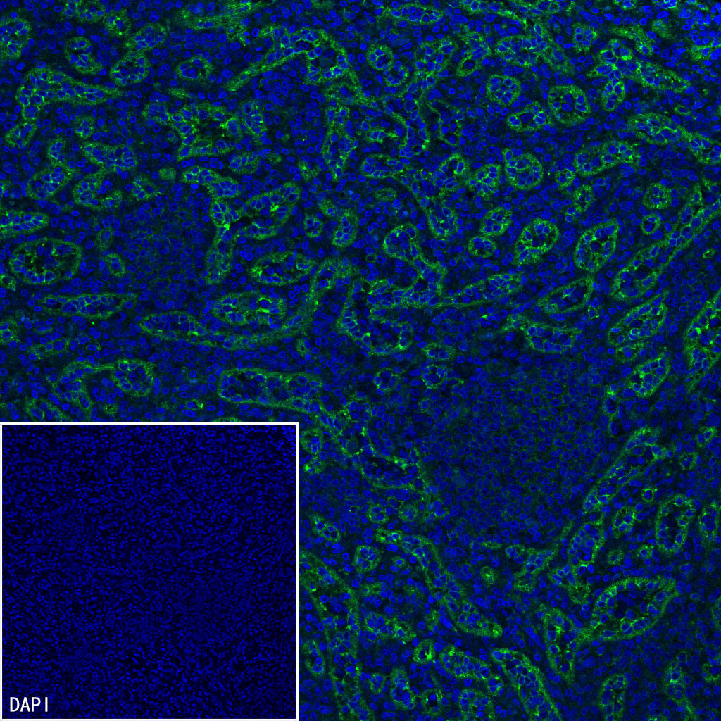
IF shows positive staining in paraffin-embedded human lung. Anti-EPCR/PROCR (CD201) antibody was used at 1/2000 dilution (Green) and incubated overnight at 4°C. Goat polyclonal Antibody to Rabbit IgG - H&L (Alexa Fluor® 488) was used as secondary antibody at 1/1000 dilution. Counterstained with DAPI (Blue). Heat mediated antigen retrieval with EDTA buffer pH9.0 was performed before commencing with IF staining protocol.
IF shows positive staining in paraffin-embedded human prostate. Anti-EPCR/PROCR (CD201) antibody was used at 1/2000 dilution (Green) and incubated overnight at 4°C. Goat polyclonal Antibody to Rabbit IgG - H&L (Alexa Fluor® 488) was used as secondary antibody at 1/1000 dilution. Counterstained with DAPI (Blue). Heat mediated antigen retrieval with EDTA buffer pH9.0 was performed before commencing with IF staining protocol.
IF shows positive staining in paraffin-embedded human spleen. Anti-EPCR/PROCR (CD201) antibody was used at 1/2000 dilution (Green) and incubated overnight at 4°C. Goat polyclonal Antibody to Rabbit IgG - H&L (Alexa Fluor® 488) was used as secondary antibody at 1/1000 dilution. Counterstained with DAPI (Blue). Heat mediated antigen retrieval with EDTA buffer pH9.0 was performed before commencing with IF staining protocol.