WB result of EPAC1 Recombinant Rabbit mAb
Primary antibody: EPAC1 Recombinant Rabbit mAb at 1/1000 dilution
Lane 1: HeLa whole cell lysate 20 µg
Lane 2: HepG2 whole cell lysate 20 µg
Lane 3: SK-OV-3 whole cell lysate 20 µg
Lane 4: HUVEC whole cell lysate 20 µg
Low expression control: HeLa whole cell lysate; HepG2 whole cell lysate
Secondary antibody: Goat Anti- rabbit IgG, (H+L), HRP conjugated at 1/10000 dilution
Predicted MW: 104 kDa
Observed MW: 95 kDa
This blot was developed with high sensitivity substrate
EPAC1 Recombinant Rabbit mAb (S-3774-58)
EPAC1 Recombinant Rabbit mAb (S-3774-58)
Price:
Regular price
$100 USD
Regular price
Sale price
$100 USD
Unit price
per
For shipping services or bulk orders, you may request a quotation.
Secure checkout with

View full details
Product Details
Product Details
Product Specification
| Host | Rabbit |
| Antigen | EPAC1 |
| Synonyms | Rap guanine nucleotide exchange factor 3; Exchange factor directly activated by cAMP 1; Exchange protein directly activated by cAMP 1 (EPAC 1); Rap1 guanine-nucleotide-exchange factor directly activated by cAMP; cAMP-regulated guanine nucleotide exchange factor I (cAMP-GEFI); CGEF1; EPAC; RAPGEF3 |
| Immunogen | Synthetic Peptide |
| Accession | O95398 |
| Clone Number | S-3774-58 |
| Antibody Type | Recombinant mAb |
| Isotype | IgG |
| Application | WB |
| Reactivity | Hu, Ms, Rt |
| Positive Sample | SK-OV-3, HUVEC, mouse brain, mouse skeletal muscle, rat brain, rat skeletal muscle |
| Purification | Protein A |
| Concentration | 0.5 mg/ml |
| Conjugation | Unconjugated |
| Physical Appearance | Liquid |
| Storage Buffer | PBS, 40% Glycerol, 0.05% BSA, 0.03% Proclin 300 |
| Stability & Storage | 12 months from date of receipt / reconstitution, -20 °C as supplied |
Dilution
| application | dilution | species |
| WB | 1:500-1:1000 | Hu, Ms, Rt |
Background
EPAC1 protein (encoded by the RAPGEF3 gene) is a guanine nucleotide exchange factor and an important effector molecule in the cAMP signaling pathway that operates independently of protein kinase A, playing a unique role in cell signal transduction and functional regulation. EPAC1 directly senses increases in intracellular second messenger cAMP concentrations through its cAMP-binding domain; upon activation, it catalyzes the conversion of the small GTPases Rap1 and Rap2 from their inactive GDP-bound state to the active GTP-bound state, thereby triggering multiple downstream signaling pathways. Unlike the classical PKA pathway, EPAC1-mediated signals play irreplaceable roles in regulating cell adhesion, intercellular junction permeability, insulin secretion, cardiac contractility, neuronal synaptic plasticity, and immune cell function. In the cardiovascular system, EPAC1 is involved in regulating endothelial cell barrier function and cardiac hypertrophy responses; in metabolic regulation, it influences glucose-stimulated insulin release; in tumor biology, abnormal expression of EPAC1 is closely associated with cancer cell migration and invasion capabilities. Furthermore, EPAC1 dysfunction is linked to various pathological conditions, including heart failure, diabetic nephropathy, chronic inflammation, and neurodegenerative diseases. Therefore, EPAC1 is not only a key nodal protein connecting cAMP signals to diverse cellular responses but has also become a novel drug target for treating cardiovascular diseases, diabetes, and cancer.
Picture
Picture
Western Blot
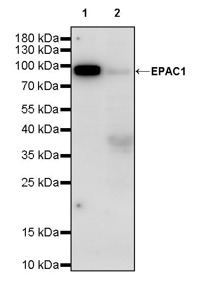
WB result of EPAC1 Recombinant Rabbit mAb
Primary antibody: EPAC1 Recombinant Rabbit mAb at 1/1000 dilution
Lane 1: mouse brain lysate 20 µg
Lane 2: mouse skeletal muscle lysate 20 µg
Secondary antibody: Goat Anti- rabbit IgG, (H+L), HRP conjugated at 1/10000 dilution
Predicted MW: 104 kDa
Observed MW: 95 kDa
This blot was developed with high sensitivity substrate
WB result of EPAC1 Recombinant Rabbit mAb
Primary antibody: EPAC1 Recombinant Rabbit mAb at 1/1000 dilution
Lane 1: rat brain lysate 20 µg
Lane 2: rat skeletal muscle lysate 20 µg
Secondary antibody: Goat Anti- rabbit IgG, (H+L), HRP conjugated at 1/10000 dilution
Predicted MW: 104 kDa
Observed MW: 95 kDa
This blot was developed with high sensitivity substrate