WB result of EDF1 Recombinant Rabbit mAb
Primary antibody: EDF1 Recombinant Rabbit mAb at 1/1000 dilution
Lane 1: Hep G2 whole cell lysate 20 μg
Lane 2: MCF7 whole cell lysate 20 μg
Lane 3: Jurkat whole cell lysate 20 μg
Lane 4: HeLa whole cell lysate 20 μg
Secondary antibody: Goat Anti-Rabbit IgG (H+L), HRP conjugated at 1/10000 dilution
Predicted MW: 16 kDa
Observed MW: 19 kDa
EDF1 Recombinant Rabbit mAb (S-4165-160)
EDF1 Recombinant Rabbit mAb (S-4165-160)
Price:
Regular price
$100 USD
Regular price
Sale price
$100 USD
Unit price
per
For shipping services or bulk orders, you may request a quotation.
Secure checkout with

View full details
Product Details
Product Details
Product Specification
| Host | Rabbit |
| Antigen | EDF1 |
| Synonyms | Endothelial differentiation-related factor 1 |
| Immunogen | Synthetic Peptide |
| Location | Cytoplasm, Nucleus |
| Accession | O60869 |
| Clone Number | S-4165-160 |
| Antibody Type | Recombinant mAb |
| Isotype | IgG |
| Application | WB, IHC-P, ICC, IF |
| Reactivity | Hu, Ms, Rt, Mk |
| Positive Sample | Hep G2, MCF7, HeLa, Jurkat, NIH/3T3, mouse brain, mouse liver, rat brain, COS-7 |
| Predicted Reactivity | Zf, Ck |
| Purification | Protein A |
| Concentration | 0.5 mg/ml |
| Conjugation | Unconjugated |
| Physical Appearance | Liquid |
| Storage Buffer | PBS, 40% Glycerol, 0.05% BSA, 0.02% sodium azide |
| Stability & Storage | 12 months from date of receipt / reconstitution, -20 °C as supplied |
Dilution
| application | dilution | species |
| WB | 1:1000 | Hu, Ms, Rt, Mk |
| IHC-P | 1:200 | Hu, Ms, Rt |
| ICC | 1:500 | Hu, Ms |
| IF | 1:500 | Ms, Rt |
Background
EDF1 (Endothelial differentiation-related factor 1, also known as multiprotein bridging factor 1/MBF1) is a 148-amino-acid basic intracellular protein encoded by the EDF1 gene that functions as a transcriptional coactivator, with its core function being to regulate transcription by bridging TATA-binding protein (TBP) and gene-specific transcription factors. It can stimulate the transcriptional activity of NR5A1 as well as ligand-dependent NR1H3/LXRA and PPARG, and enhances the DNA-binding capacity of ATF1, ATF2, CREB1, and NR5A1. EDF1 shuttles between the cytoplasm and the nucleus; its IQ motif overlaps with the binding regions for nuclear receptors and transcription factors, and phosphorylation may serve as a switch between these two activities. In the cytoplasm, it may also regulate nitric oxide synthase activity by sequestering calmodulin. The protein is most abundantly expressed in adult heart, liver, and pancreas, as well as in fetal kidney, and is presumed to participate in endothelial cell differentiation, hormone-induced cardiomyocyte hypertrophy, and lipid metabolism, among other processes.
Picture
Picture
Western Blot
WB result of EDF1 Recombinant Rabbit mAb
Primary antibody: EDF1 Recombinant Rabbit mAb at 1/1000 dilution
Lane 1: NIH/3T3 whole cell lysate 20 μg
Lane 2: mouse brain lysate 20 μg
Lane 3: mouse liver lysate 20 μg
Secondary antibody: Goat Anti-Rabbit IgG (H+L), HRP conjugated at 1/10000 dilution
Predicted MW: 16 kDa
Observed MW: 19 kDa
WB result of EDF1 Recombinant Rabbit mAb
Primary antibody: EDF1 Recombinant Rabbit mAb at 1/1000 dilution
Lane 1: rat brain lysate 20 μg
Secondary antibody: Goat Anti-Rabbit IgG (H+L), HRP conjugated at 1/10000 dilution
Predicted MW: 16 kDa
Observed MW: 19 kDa
WB result of EDF1 Recombinant Rabbit mAb
Primary antibody: EDF1 Recombinant Rabbit mAb at 1/1000 dilution
Lane 1: COS-7 whole cell lysate 20 μg
Secondary antibody: Goat Anti-Rabbit IgG (H+L), HRP conjugated at 1/10000 dilution
Predicted MW: 16 kDa
Observed MW: 19 kDa
Immunohistochemistry
IHC shows positive staining in paraffin-embedded human brain. Anti-EDF1 antibody was used at 1/200 dilution, followed by a HRP Polymer for Rabbit IgG (ready to use). Counterstained with hematoxylin. Heat mediated antigen retrieval with Tris/EDTA buffer pH9.0 was performed before commencing with IHC staining protocol.
IHC shows positive staining in paraffin-embedded human stomach. Anti-EDF1 antibody was used at 1/200 dilution, followed by a HRP Polymer for Rabbit IgG (ready to use). Counterstained with hematoxylin. Heat mediated antigen retrieval with Tris/EDTA buffer pH9.0 was performed before commencing with IHC staining protocol.
IHC shows positive staining in paraffin-embedded human breast cancer. Anti-EDF1 antibody was used at 1/200 dilution, followed by a HRP Polymer for Rabbit IgG (ready to use). Counterstained with hematoxylin. Heat mediated antigen retrieval with Tris/EDTA buffer pH9.0 was performed before commencing with IHC staining protocol.
IHC shows positive staining in paraffin-embedded mouse kidney. Anti-EDF1 antibody was used at 1/200 dilution, followed by a HRP Polymer for Rabbit IgG (ready to use). Counterstained with hematoxylin. Heat mediated antigen retrieval with Tris/EDTA buffer pH9.0 was performed before commencing with IHC staining protocol.
IHC shows positive staining in paraffin-embedded rat kidney. Anti-EDF1 antibody was used at 1/200 dilution, followed by a HRP Polymer for Rabbit IgG (ready to use). Counterstained with hematoxylin. Heat mediated antigen retrieval with Tris/EDTA buffer pH9.0 was performed before commencing with IHC staining protocol.
Immunocytochemistry
ICC shows positive staining in HeLa cells. Anti-EDF1 antibody was used at 1/500 dilution (Green) and incubated overnight at 4°C. Goat polyclonal Antibody to Rabbit IgG - H&L (Alexa Fluor® 488) was used as secondary antibody at 1/1000 dilution. The cells were fixed with 4% PFA and permeabilized with 0.1% PBS-Triton X-100. Nuclei were counterstained with DAPI (Blue). Counterstain with tubulin (Red).
ICC shows positive staining in NIH/3T3 cells. Anti-EDF1 antibody was used at 1/500 dilution (Green) and incubated overnight at 4°C. Goat polyclonal Antibody to Rabbit IgG - H&L (Alexa Fluor® 488) was used as secondary antibody at 1/1000 dilution. The cells were fixed with 4% PFA and permeabilized with 0.1% PBS-Triton X-100. Nuclei were counterstained with DAPI (Blue). Counterstain with tubulin (Red).
Immunofluorescence
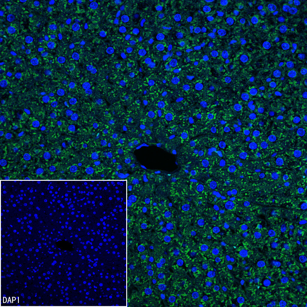
IF shows positive staining in paraffin-embedded mouse liver. Anti-EDF1 antibody was used at 1/500 dilution (Green) and incubated overnight at 4°C. Goat polyclonal Antibody to Rabbit IgG - H&L (Alexa Fluor® 488) was used as secondary antibody at 1/1000 dilution. Counterstained with DAPI (Blue). Heat mediated antigen retrieval with EDTA buffer pH9.0 was performed before commencing with IF staining protocol.
IF shows positive staining in paraffin-embedded rat liver. Anti-EDF1 antibody was used at 1/500 dilution (Green) and incubated overnight at 4°C. Goat polyclonal Antibody to Rabbit IgG - H&L (Alexa Fluor® 488) was used as secondary antibody at 1/1000 dilution. Counterstained with DAPI (Blue). Heat mediated antigen retrieval with EDTA buffer pH9.0 was performed before commencing with IF staining protocol.