WB result of Cytokeratin 7 Recombinant Rabbit mAb
Primary antibody: Cytokeratin 7 Recombinant Rabbit mAb at 1/5000 dilution
Lane 1: MCF7 whole cell lysate 20 µg
Lane 2: A549 whole cell lysate 20 µg
Lane 3: Hela whole cell lysate 20 µg
Lane 4: SK-BR-3 whole cell lysate 20 µg
Lane 5: T-47D whole cell lysate 20 µg
Negative control: MCF7 whole cell lysate
Secondary antibody: Goat Anti-Rabbit IgG (H+L), HRP conjugated at 1/10000 dilution
Predicted MW: 51 kDa
Observed MW: 38-55 kDa
Cytokeratin 7 Recombinant Rabbit mAb (S-5954)
Cytokeratin 7 Recombinant Rabbit mAb (S-5954)
Price:
Regular price
$100 USD
Regular price
Sale price
$100 USD
Unit price
per
For shipping services or bulk orders, you may request a quotation.
Secure checkout with

View full details
Product Details
Product Details
Product Specification
| Host | Rabbit |
| Antigen | Cytokeratin 7 |
| Location | Cytoplasm |
| Accession | P08729 |
| Clone Number | S-5954 |
| Antibody Type | Recombinant mAb |
| Isotype | IgG |
| Application | WB, IHC-P, ICC, IF |
| Reactivity | Hu, Ms, Rt |
| Positive Sample | A549, Hela, SK-BR-3, T-47D, mouse kidney, mouse lung, rat kidney, rat lung, rat liver |
| Purification | Protein A |
| Concentration | 0.25 mg/ml |
| Conjugation | Unconjugated |
| Physical Appearance | Liquid |
| Storage Buffer | PBS, 40% Glycerol, 0.05% BSA, 0.02% sodium azide |
| Stability & Storage | 12 months from date of receipt / reconstitution, -20 °C as supplied |
Dilution
| application | dilution | species |
| WB | 1:1000-1:10000 | Hu, Ms, Rt |
| IHC-P | 1:2000 | Hu, Ms, Rt |
| ICC | 1:250 | Hu |
| IF | 1:250 | Ms, Rt |
Background
Cytokeratin 7 (CK7), also known as keratin 7 (KRT7), is a type II basic low-molecular-weight cytokeratin encoded by the KRT7 gene located on chromosome 12q13.13, and it is expressed in simple epithelia of various organs, including the female genital tract, lung, breast, and urinary tract. As an intermediate filament protein, CK7 forms heterotetramers with type I keratins and functions to maintain the structural integrity of epithelial cells. In diagnostic pathology, CK7 is widely used as an immunohistochemical marker, and its expression pattern—often evaluated in combination with CK20—is instrumental in determining the site of origin for metastatic adenocarcinomas, such as distinguishing primary lung adenocarcinomas from colorectal metastases. Beyond its diagnostic utility, CK7 expression has been associated with prognostic significance in several malignancies, including worse outcomes in pulmonary large-cell neuroendocrine carcinoma, cervical adenocarcinoma, and ovarian cancer. Recent molecular studies have also identified CK7 as a key effector in cancer progression, with N6-methyladenosine (m6A) modification of KRT7 mRNA promoting breast cancer lung metastasis, and its involvement in epithelial-mesenchymal transition in ovarian cancer.
Picture
Picture
Western Blot
WB result of Cytokeratin 7 Recombinant Rabbit mAb
Primary antibody: Cytokeratin 7 Recombinant Rabbit mAb at 1/5000 dilution
Lane 1: mouse kidney lysate 20 µg
Lane 2: mouse lung lysate 20 µg
Secondary antibody: Goat Anti-Rabbit IgG (H+L), HRP conjugated at 1/10000 dilution
Predicted MW: 51 kDa
Observed MW: 38-55 kDa
WB result of Cytokeratin 7 Recombinant Rabbit mAb
Primary antibody: Cytokeratin 7 Recombinant Rabbit mAb at 1/5000 dilution
Lane 1: rat kidney lysate 20 µg
Lane 2: rat liver lysate 20 µg
Lane 3: rat lung lysate 20 µg
Secondary antibody: Goat Anti-Rabbit IgG (H+L), HRP conjugated at 1/10000 dilution
Predicted MW: 51 kDa
Observed MW: 36-50 kDa
Immunohistochemistry
IHC shows positive staining in paraffin-embedded human breast. Anti-Cytokeratin 7 antibody was used at 1/2000 dilution, followed by a HRP Polymer for Rabbit IgG (ready to use). Counterstained with hematoxylin. Heat mediated antigen retrieval with Tris/EDTA buffer pH9.0 was performed before commencing with IHC staining protocol.
IHC shows positive staining in paraffin-embedded human breast cancer. Anti-Cytokeratin 7 antibody was used at 1/2000 dilution, followed by a HRP Polymer for Rabbit IgG (ready to use). Counterstained with hematoxylin. Heat mediated antigen retrieval with Tris/EDTA buffer pH9.0 was performed before commencing with IHC staining protocol.
IHC shows positive staining in paraffin-embedded human bladder cancer. Anti-Cytokeratin 7 antibody was used at 1/2000 dilution, followed by a HRP Polymer for Rabbit IgG (ready to use). Counterstained with hematoxylin. Heat mediated antigen retrieval with Tris/EDTA buffer pH9.0 was performed before commencing with IHC staining protocol.
IHC shows positive staining in paraffin-embedded human ovarian cancer. Anti-Cytokeratin 7 antibody was used at 1/2000 dilution, followed by a HRP Polymer for Rabbit IgG (ready to use). Counterstained with hematoxylin. Heat mediated antigen retrieval with Tris/EDTA buffer pH9.0 was performed before commencing with IHC staining protocol.
Negative control: IHC shows negative staining in paraffin-embedded human renal cell carcinoma. Anti-Cytokeratin 7 antibody was used at 1/2000 dilution, followed by a HRP Polymer for Rabbit IgG (ready to use). Counterstained with hematoxylin. Heat mediated antigen retrieval with Tris/EDTA buffer pH9.0 was performed before commencing with IHC staining protocol.
IHC shows positive staining in paraffin-embedded mouse kidney. Anti-Cytokeratin 7 antibody was used at 1/2000 dilution, followed by a HRP Polymer for Rabbit IgG (ready to use). Counterstained with hematoxylin. Heat mediated antigen retrieval with Tris/EDTA buffer pH9.0 was performed before commencing with IHC staining protocol.
IHC shows positive staining in paraffin-embedded mouse liver. Anti-Cytokeratin 7 antibody was used at 1/2000 dilution, followed by a HRP Polymer for Rabbit IgG (ready to use). Counterstained with hematoxylin. Heat mediated antigen retrieval with Tris/EDTA buffer pH9.0 was performed before commencing with IHC staining protocol.
IHC shows positive staining in paraffin-embedded rat kidney. Anti-Cytokeratin 7 antibody was used at 1/2000 dilution, followed by a HRP Polymer for Rabbit IgG (ready to use). Counterstained with hematoxylin. Heat mediated antigen retrieval with Tris/EDTA buffer pH9.0 was performed before commencing with IHC staining protocol.
Immunocytochemistry
ICC shows positive staining in A549 cells. Anti- Cytokeratin 7 antibody was used at 1/250 dilution (Green) and incubated overnight at 4°C. Goat polyclonal Antibody to Rabbit IgG - H&L (Alexa Fluor® 488) was used as secondary antibody at 1/1000 dilution. The cells were fixed with 100% ice-cold methanol and permeabilized with 0.1% PBS-Triton X-100. Nuclei were counterstained with DAPI (Blue). Counterstain with tubulin (Red).
Immunofluorescence
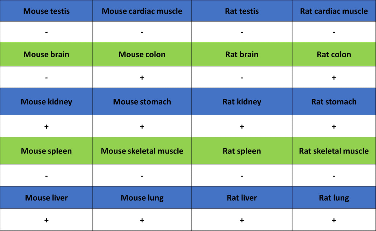
IF shows positive staining in paraffin-embedded mouse colon. Anti- Cytokeratin 7 antibody was used at 1/250 dilution (Green) and incubated overnight at 4°C. Goat polyclonal Antibody to Rabbit IgG - H&L (Alexa Fluor® 488) was used as secondary antibody at 1/1000 dilution. Counterstained with DAPI (Blue). Heat mediated antigen retrieval with EDTA buffer pH9.0 was performed before commencing with IF staining protocol.
IF shows positive staining in paraffin-embedded mouse kidney. Anti- Cytokeratin 7 antibody was used at 1/250 dilution (Green) and incubated overnight at 4°C. Goat polyclonal Antibody to Rabbit IgG - H&L (Alexa Fluor® 488) was used as secondary antibody at 1/1000 dilution. Counterstained with DAPI (Blue). Heat mediated antigen retrieval with EDTA buffer pH9.0 was performed before commencing with IF staining protocol.
IF shows positive staining in paraffin-embedded mouse stomach. Anti- Cytokeratin 7 antibody was used at 1/250 dilution (Green) and incubated overnight at 4°C. Goat polyclonal Antibody to Rabbit IgG - H&L (Alexa Fluor® 488) was used as secondary antibody at 1/1000 dilution. Counterstained with DAPI (Blue). Heat mediated antigen retrieval with EDTA buffer pH9.0 was performed before commencing with IF staining protocol.
IF shows positive staining in paraffin-embedded rat colon. Anti- Cytokeratin 7 antibody was used at 1/250 dilution (Green) and incubated overnight at 4°C. Goat polyclonal Antibody to Rabbit IgG - H&L (Alexa Fluor® 488) was used as secondary antibody at 1/1000 dilution. Counterstained with DAPI (Blue). Heat mediated antigen retrieval with EDTA buffer pH9.0 was performed before commencing with IF staining protocol.
IF shows positive staining in paraffin-embedded rat kidney. Anti- Cytokeratin 7 antibody was used at 1/250 dilution (Green) and incubated overnight at 4°C. Goat polyclonal Antibody to Rabbit IgG - H&L (Alexa Fluor® 488) was used as secondary antibody at 1/1000 dilution. Counterstained with DAPI (Blue). Heat mediated antigen retrieval with EDTA buffer pH9.0 was performed before commencing with IF staining protocol.
IF shows positive staining in paraffin-embedded rat stomach. Anti- Cytokeratin 7 antibody was used at 1/250 dilution (Green) and incubated overnight at 4°C. Goat polyclonal Antibody to Rabbit IgG - H&L (Alexa Fluor® 488) was used as secondary antibody at 1/1000 dilution. Counterstained with DAPI (Blue). Heat mediated antigen retrieval with EDTA buffer pH9.0 was performed before commencing with IF staining protocol.