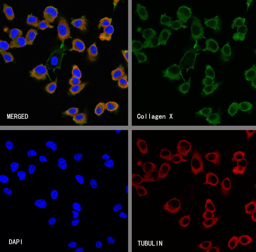
ICC shows positive staining in HT-1080 cells. Anti-Collagen X antibody was used at 1/1000 dilution (Green) and incubated overnight at 4°C. Goat polyclonal Antibody to Rabbit IgG - H&L (Alexa Fluor® 488) was used as secondary antibody at 1/1000 dilution. The cells were fixed with 100% ice-cold methanol and permeabilized with 0.1% PBS-Triton X-100. Nuclei were counterstained with DAPI (Blue). Counterstain with tubulin (Red).
Collagen X Recombinant Mouse mAb (S-M0004)
Collagen X Recombinant Mouse mAb (S-M0004)
Price:
Regular price
$100 USD
Regular price
Sale price
$100 USD
Unit price
per
For shipping services or bulk orders, you may request a quotation.
Secure checkout with

View full details
Product Details
Product Details
Product Specification
| Host | Mouse |
| Antigen | Collagen X |
| Synonyms | Collagen alpha-1(X) chain; COL10A1 |
| Location | Secreted |
| Accession | Q03692 |
| Clone Number | S-M0004 |
| Antibody Type | Mouse mAb |
| Isotype | IgM |
| Application | IHC-P, ICC |
| Reactivity | Hu |
| Concentration | 1 mg/ml |
| Conjugation | Unconjugated |
| Physical Appearance | Liquid |
| Storage Buffer | PBS, 40% Glycerol, 0.05% BSA, 0.02% sodium azide |
| Stability & Storage | 12 months from date of receipt / reconstitution, -20 °C as supplied |
Dilution
| application | dilution | species |
| IHC-P | 1:1000 | Hu |
| ICC | 1:1000 | Hu |
Background
Collagen X, also known as COL10A1, is a short-chain, non-fibril-forming network-forming collagen belonging to the MSC (membrane-associated collagen) family, which forms a homotrimer composed of three identical α1(X) chains. Its key distinction from other collagens lies in its specific synthesis and secretion by hypertrophic chondrocytes, making it a hallmark molecule of extracellular matrix remodeling during endochondral ossification. Within the growth plate, Collagen X does not constitute the main structural framework supporting cartilage architecture, as type II collagen does; instead, it is concentrated in the pericellular matrix of hypertrophic chondrocytes and at the interface between the growth plate and bone tissue (i.e., the calcified cartilage zone). Its function is to provide a structural scaffold for the cartilage matrix that is destined to be replaced by bone tissue, promote the release of matrix vesicles to initiate hydroxyapatite deposition, and thereby participate in the calcification process. From a pathological perspective, abnormal expression of Collagen X is a core link in various skeletal development disorders: loss-of-function mutations in the COL10A1 gene can lead to metaphyseal chondrodysplasia (Schmid type, MCDS), characterized by disorganized hypertrophic zones, failed calcification, and impaired trabecular bone formation in the metaphysis, with clinical features including short-limbed dwarfism and coxa vara. Furthermore, in degenerative joint diseases such as osteoarthritis, articular chondrocytes that normally do not express Collagen X undergo dedifferentiation under pathological stimulation, reactivate the hypertrophic program, and ectopically express Collagen X—a process considered an important molecular hallmark of articular cartilage degeneration and calcification. Therefore, Collagen X serves both as a physiological marker of skeletal development and growth plate mineralization and as a pathological marker of chondrocyte hypertrophy, holding significant diagnostic and prognostic value in developmental biology and joint disease research.
Picture
Picture
Immunocytochemistry