IHC shows positive staining in paraffin-embedded human liver. Anti-Coagulation factor V antibody was used at 1/500 dilution, followed by a HRP Polymer for Rabbit IgG (ready to use). Counterstained with hematoxylin. Heat mediated antigen retrieval with Tris/EDTA buffer pH9.0 was performed before commencing with IHC staining protocol.
Coagulation factor V Recombinant Rabbit mAb (S-4489-58)
Coagulation factor V Recombinant Rabbit mAb (S-4489-58)
Price:
Regular price
$100 USD
Regular price
Sale price
$100 USD
Unit price
per
For shipping services or bulk orders, you may request a quotation.
Secure checkout with

View full details
Product Details
Product Details
Product Specification
| Host | Rabbit |
| Antigen | Coagulation factor V |
| Synonyms | Activated protein C cofactor; Proaccelerin; labile factor; F5 |
| Immunogen | Synthetic Peptide |
| Location | Secreted |
| Accession | P12259 |
| Clone Number | S-4489-58 |
| Antibody Type | Recombinant mAb |
| Isotype | IgG |
| Application | IHC-P, IF |
| Reactivity | Hu, Ms, Rt |
| Predicted Reactivity | Pg |
| Purification | Protein A |
| Concentration | 2 mg/ml |
| Conjugation | Unconjugated |
| Physical Appearance | Liquid |
| Storage Buffer | PBS, 40% Glycerol, 0.05% BSA, 0.02% sodium azide |
| Stability & Storage | 12 months from date of receipt / reconstitution, -20 °C as supplied |
Dilution
| application | dilution | species |
| IHC-P | 1:500 | Hu, Ms, Rt |
| IF | 1:2000 | Ms, Rt |
Background
Coagulation factor V (FV) is a high-molecular-weight glycoprotein synthesized in the liver and present in plasma in an inactive form, serving as an indispensable and critical cofactor in the coagulation cascade. During the coagulation process, it is proteolytically cleaved by thrombin or factor Xa (FXa) through limited proteolysis to generate activated factor Va (FVa), which then binds to activated factor X (FXa) on platelet phospholipid surfaces in a 1:1 stoichiometric ratio, forming a molecular machine known as the "prothrombinase complex." This complex efficiently converts prothrombin to thrombin, thereby initiating an explosive amplification of the coagulation cascade and ultimately promoting the conversion of fibrinogen to fibrin to form a stable thrombus. Beyond its classical procoagulant role in hemostasis, FV also exerts anticoagulant effects through its splicing isoforms (such as FV-short), which act as a cofactor for tissue factor pathway inhibitor α (TFPIα) to negatively regulate the initiation of coagulation by inhibiting the tissue factor pathway, illustrating its "dual-faced" nature in hemostatic balance. Mutations in the FV gene give rise to a range of clinical disorders: for instance, the FV Leiden mutation (R506Q) confers resistance to proteolytic inactivation by activated protein C (APC) and is the most common inherited cause of thrombophilia in Caucasian populations; whereas severe FV deficiency (parhemophilia) is a rare autosomal recessive bleeding disorder with variable bleeding symptoms, and treatment typically requires transfusion of fresh frozen plasma or platelets, as FV is present in both plasma and platelet α-granules, and the two pools are not fully interchangeable in their hemostatic functions.
Picture
Picture
Immunohistochemistry
Negative control: IHC shows negative staining in paraffin-embedded human brain. Anti-Coagulation factor V antibody was used at 1/500 dilution, followed by a HRP Polymer for Rabbit IgG (ready to use). Counterstained with hematoxylin. Heat mediated antigen retrieval with Tris/EDTA buffer pH9.0 was performed before commencing with IHC staining protocol.
Negative control: IHC shows negative staining in paraffin-embedded human colon cancer. Anti-Coagulation factor V antibody was used at 1/500 dilution, followed by a HRP Polymer for Rabbit IgG (ready to use). Counterstained with hematoxylin. Heat mediated antigen retrieval with Tris/EDTA buffer pH9.0 was performed before commencing with IHC staining protocol.
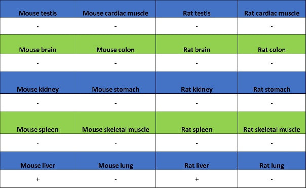
IHC shows positive staining in paraffin-embedded mouse liver. Anti-Coagulation factor V antibody was used at 1/500 dilution, followed by a HRP Polymer for Rabbit IgG (ready to use). Counterstained with hematoxylin. Heat mediated antigen retrieval with Tris/EDTA buffer pH9.0 was performed before commencing with IHC staining protocol.
Negative control: IHC shows negative staining in paraffin-embedded mouse brain. Anti-Coagulation factor V antibody was used at 1/500 dilution, followed by a HRP Polymer for Rabbit IgG (ready to use). Counterstained with hematoxylin. Heat mediated antigen retrieval with Tris/EDTA buffer pH9.0 was performed before commencing with IHC staining protocol.
IHC shows positive staining in paraffin-embedded rat liver. Anti-Coagulation factor V antibody was used at 1/500 dilution, followed by a HRP Polymer for Rabbit IgG (ready to use). Counterstained with hematoxylin. Heat mediated antigen retrieval with Tris/EDTA buffer pH9.0 was performed before commencing with IHC staining protocol.
Negative control: IHC shows negative staining in paraffin-embedded rat colon. Anti-Coagulation factor V antibody was used at 1/500 dilution, followed by a HRP Polymer for Rabbit IgG (ready to use). Counterstained with hematoxylin. Heat mediated antigen retrieval with Tris/EDTA buffer pH9.0 was performed before commencing with IHC staining protocol.
Immunofluorescence
IF shows positive staining in paraffin-embedded mouse liver. Anti- Coagulation factor V antibody was used at 1/2000 dilution (Green) and incubated overnight at 4°C. Goat polyclonal Antibody to Rabbit IgG - H&L (Alexa Fluor® 488) was used as secondary antibody at 1/1000 dilution. Counterstained with DAPI (Blue). Heat mediated antigen retrieval with EDTA buffer pH9.0 was performed before commencing with IF staining protocol.
Negative control: IF shows negative staining in paraffin-embedded mouse brain. Anti-Coagulation factor V antibody was used at 1/2000 dilution and incubated overnight at 4°C. Goat polyclonal Antibody to Rabbit IgG - H&L (Alexa Fluor® 488) was used as secondary antibody at 1/1000 dilution. Counterstained with DAPI (Blue). Heat mediated antigen retrieval with EDTA buffer pH9.0 was performed before commencing with IF staining protocol.
IF shows positive staining in paraffin-embedded rat liver. Anti- Coagulation factor V antibody was used at 1/2000 dilution (Green) and incubated overnight at 4°C. Goat polyclonal Antibody to Rabbit IgG - H&L (Alexa Fluor® 488) was used as secondary antibody at 1/1000 dilution. Counterstained with DAPI (Blue). Heat mediated antigen retrieval with EDTA buffer pH9.0 was performed before commencing with IF staining protocol.
Negative control: IF shows negative staining in paraffin-embedded rat brain. Anti- Coagulation factor V antibody was used at 1/2000 dilution and incubated overnight at 4°C. Goat polyclonal Antibody to Rabbit IgG - H&L (Alexa Fluor® 488) was used as secondary antibody at 1/1000 dilution. Counterstained with DAPI (Blue). Heat mediated antigen retrieval with EDTA buffer pH9.0 was performed before commencing with IF staining protocol.