WB result of Claudin 3+Claudin 9 Recombinant Rabbit mAb
Primary antibody: Claudin 3+Claudin 9 Recombinant Rabbit mAb at 1/1000 dilution
Lane 1: COLO 205 whole cell lysate 20 µg
Lane 2: SW480 whole cell lysate 20 µg
Lane 3: MCF7 whole cell lysate 20 µg
Lane 4: A431 whole cell lysate 20 µg
Secondary antibody: Goat Anti-rabbit IgG, (H+L), HRP conjugated at 1/10000 dilution
Predicted MW: 23 kDa
Observed MW: 18 kDa
Claudin 3+Claudin 9 Recombinant Rabbit mAb (SDT-1452-80)
Claudin 3+Claudin 9 Recombinant Rabbit mAb (SDT-1452-80)
Price:
Regular price
$100 USD
Regular price
Sale price
$100 USD
Unit price
per
For shipping services or bulk orders, you may request a quotation.
Secure checkout with

View full details
Product Details
Product Details
Product Specification
| Host | Rabbit |
| Synonyms | CLDN9 |
| Immunogen | Synthetic Peptide |
| Location | Cell membrane |
| Accession | O95484 |
| Clone Number | SDT-1452-80 |
| Antibody Type | Recombinant mAb |
| Isotype | IgG |
| Application | WB, IHC-P, ICC, ICFCM |
| Reactivity | Hu, Rt |
| Predicted Reactivity | Ms |
| Purification | Protein A |
| Concentration | 0.5 mg/ml |
| Conjugation | Unconjugated |
| Physical Appearance | Liquid |
| Storage Buffer | PBS, 40% Glycerol, 0.05% BSA, 0.03% Proclin 300 |
| Stability & Storage | 12 months from date of receipt / reconstitution, -20 °C as supplied |
Dilution
| application | dilution | species |
| WB | 1:1000 | Hu |
| IHC-P | 1:500 | Hu |
| ICC | 1:100 | Hu |
| ICFCM | 1:50 | Hu |
Background
Claudin-3 and Claudin-9 are two closely related members of the Claudin family of tight junction proteins, both belonging to the classical Claudin subclass that primarily serves barrier functions. Their most notable shared features lie in their highly similar protein sequences and common receptor properties: Claudin-3 and Claudin-9 share a consistent overall fold and both can bind Clostridium perfringens enterotoxin (CPE). In terms of expression and distribution, they display patterns that are both overlapping and distinct: Claudin-3 is highly expressed in choroid plexus epithelium, where it constitutes an important component of the blood-cerebrospinal fluid barrier, and is also commonly found in various epithelia such as the ovary and mammary gland; Claudin-9, on the other hand, has been identified in the inner ear, cornea, liver, and developing kidney, and serves as one of the auxiliary receptors for hepatitis C virus. Regarding physiological function, both contribute to enhancing epithelial barrier tightness, but their specific roles differ: Claudin-3 expression declines during development, whereas Claudin-9 expression increases with development, suggesting that they undertake distinct tasks during barrier maturation. Furthermore, Claudin-3 is dysregulated in multiple cancers and is often regarded as a potential therapeutic target, while Claudin-9 has also been proposed to possibly participate in the formation of tricellular junction barriers.
Picture
Picture
Western Blot
WB result of Claudin 3+Claudin 9 Recombinant Rabbit mAb
Primary antibody: Claudin 3+Claudin 9 Recombinant Rabbit mAb at 1/1000 dilution
Lane 1: PC-12 whole cell lysate 20 µg
Secondary antibody: Goat Anti-rabbit IgG, (H+L), HRP conjugated at 1/10000 dilution
Predicted MW: 23 kDa
Observed MW: 18 kDa
FC
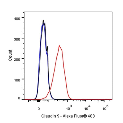
Flow cytometric analysis of 4% PFA fixed 90% methanol permeabilized COLO 205 (Human colon cancer cell) labelling anti-Claudin 3+Claudin 9 antibody at 1/50 dilution (1 μg) / (Red) compared with a Rabbit monoclonal IgG (Black) isotype control and an unlabelled control (cells without incubation with primary antibody and secondary antibody) (Blue). Goat Anti - Rabbit IgG Alexa Fluor® 488 was used as the secondary antibody.
Immunohistochemistry
IHC shows positive staining in paraffin-embedded human colon. Anti-Claudin 3+Claudin 9 antibody was used at 1/500 dilution, followed by a HRP Polymer for Mouse & Rabbit IgG (ready to use). Counterstained with hematoxylin. Heat mediated antigen retrieval with Tris/EDTA buffer pH9.0 was performed before commencing with IHC staining protocol.
IHC shows positive staining in paraffin-embedded human tonsil. Anti-Claudin 3+Claudin 9 antibody was used at 1/500 dilution, followed by a HRP Polymer for Mouse & Rabbit IgG (ready to use). Counterstained with hematoxylin. Heat mediated antigen retrieval with Tris/EDTA buffer pH9.0 was performed before commencing with IHC staining protocol.
IHC shows positive staining in paraffin-embedded human stomach. Anti-Claudin 3+Claudin 9 antibody was used at 1/500 dilution, followed by a HRP Polymer for Mouse & Rabbit IgG (ready to use). Counterstained with hematoxylin. Heat mediated antigen retrieval with Tris/EDTA buffer pH9.0 was performed before commencing with IHC staining protocol.
Negative control: IHC shows negative staining in paraffin-embedded human breast. Anti- Claudin 3+Claudin 9 antibody was used at 1/500 dilution, followed by a HRP Polymer for Mouse & Rabbit IgG (ready to use). Counterstained with hematoxylin. Heat mediated antigen retrieval with Tris/EDTA buffer pH9.0 was performed before commencing with IHC staining protocol.
IHC shows positive staining in paraffin-embedded human breast cancer. Anti-Claudin 3+Claudin 9 antibody was used at 1/500 dilution, followed by a HRP Polymer for Mouse & Rabbit IgG (ready to use). Counterstained with hematoxylin. Heat mediated antigen retrieval with Tris/EDTA buffer pH9.0 was performed before commencing with IHC staining protocol.
IHC shows positive staining in paraffin-embedded human colon cancer. Anti-Claudin 3+Claudin 9 antibody was used at 1/500 dilution, followed by a HRP Polymer for Mouse & Rabbit IgG (ready to use). Counterstained with hematoxylin. Heat mediated antigen retrieval with Tris/EDTA buffer pH9.0 was performed before commencing with IHC staining protocol.
IHC shows positive staining in paraffin-embedded human ovarian cancer. Anti- Claudin 3+Claudin 9 antibody was used at 1/500 dilution, followed by a HRP Polymer for Mouse & Rabbit IgG (ready to use). Counterstained with hematoxylin. Heat mediated antigen retrieval with Tris/EDTA buffer pH9.0 was performed before commencing with IHC staining protocol.
IHC shows positive staining in paraffin-embedded human prostatic cancer. Anti- Claudin 3+Claudin 9 antibody was used at 1/500 dilution, followed by a HRP Polymer for Mouse & Rabbit IgG (ready to use). Counterstained with hematoxylin. Heat mediated antigen retrieval with Tris/EDTA buffer pH9.0 was performed before commencing with IHC staining protocol.
IHC shows positive staining in paraffin-embedded human gastric cancer. Anti-Claudin 3+Claudin 9 antibody was used at 1/500 dilution, followed by a HRP Polymer for Mouse & Rabbit IgG (ready to use). Counterstained with hematoxylin. Heat mediated antigen retrieval with Tris/EDTA buffer pH9.0 was performed before commencing with IHC staining protocol.
Immunocytochemistry
ICC shows positive staining in COLO 205 cells. Anti-Claudin 3+Claudin 9 antibody was used at 1/100 dilution (Green) and incubated overnight at 4°C. Goat polyclonal Antibody to Rabbit IgG - H&L (Alexa Fluor® 488) was used as secondary antibody at 1/1000 dilution. The cells were fixed with 4% PFA and permeabilized with 0.1% PBS-Triton X-100. Nuclei were counterstained with DAPI (Blue). Counterstain with tubulin (Red).