WB result of Choline Kinase α/CHKA Recombinant Rabbit mAb
Primary antibody: Choline Kinase α/CHKA Recombinant Rabbit mAb at 1/1000 dilution
Lane 1: Jurkat whole cell lysate 20 µg
Lane 2: HepG2 whole cell lysate 20 µg
Lane 3: MCF7 whole cell lysate 20 µg
Lane 4: SK-BR-3 whole cell lysate 20 µg
Secondary antibody: Goat Anti-Rabbit IgG, (H+L), HRP conjugated at 1/10000 dilution
Predicted MW: 52 kDa
Observed MW: 50 kDa
Choline Kinase α/CHKA Recombinant Rabbit mAb (S-4422-31)
Choline Kinase α/CHKA Recombinant Rabbit mAb (S-4422-31)
Price:
Regular price
$100 USD
Regular price
Sale price
$100 USD
Unit price
per
For shipping services or bulk orders, you may request a quotation.
Secure checkout with

View full details
Product Details
Product Details
Product Specification
| Host | Rabbit |
| Antigen | Choline Kinase α/CHKA |
| Synonyms | Choline kinase alpha; CHETK-alpha; Ethanolamine kinase (EK); CHK; CKI |
| Immunogen | Synthetic Peptide |
| Location | Cytoplasm |
| Accession | P35790 |
| Clone Number | S-4422-31 |
| Antibody Type | Recombinant mAb |
| Isotype | IgG |
| Application | WB, IHC-P |
| Reactivity | Hu, Mk |
| Positive Sample | Jurkat, HepG2, MCF7, SK-BR-3, COS7 |
| Purification | Protein A |
| Concentration | 0.5 mg/ml |
| Conjugation | Unconjugated |
| Physical Appearance | Liquid |
| Storage Buffer | PBS, 40% Glycerol, 0.05% BSA, 0.02% sodium azide |
| Stability & Storage | 12 months from date of receipt / reconstitution, -20 °C as supplied |
Dilution
| application | dilution | species |
| WB | 1:1000 | Hu, Mk |
| IHC-P | 1:1000 | Hu |
Background
Choline kinase alpha (CHKA or ChoKα) is the first key enzyme in the Kennedy pathway of phosphatidylcholine biosynthesis, catalyzing the phosphorylation of free choline to phosphocholine in the cytoplasm, a step essential for maintaining cell membrane structural integrity and supporting cell proliferation. The human gene encoding CHKA is located on chromosome 11q13.2, spans approximately 69 kb, and through alternative splicing produces two isoforms of 457 and 439 amino acid residues; its active form functions as a homo- or heterodimer and requires ATP and Mg²⁺ for catalytic activity. Structural biology studies have revealed that its active center contains an ATP-binding pocket formed by two lobes and a deep hydrophobic groove in the C-terminal domain serving as the choline-binding site, and it exhibits a unique double-displacement catalytic mechanism in which the γ-phosphate group of ATP is first transferred to the enzyme itself to form a phosphorylated enzyme intermediate, which then transfers the phosphate to choline. In addition to catalyzing choline phosphorylation, CHKA also retains ethanolamine kinase activity, capable of phosphorylating ethanolamine and participating in phosphatidylethanolamine synthesis. In cancer biology, CHKA is considered an important oncogene, overexpressed in approximately 40–60% of human tumors (such as breast cancer, lung cancer, colorectal cancer, and glioma), and its high expression is closely associated with poor patient prognosis. Its oncogenic mechanisms not only involve providing phospholipid precursors for the robust membrane synthesis of tumor cells but also include its enzymatic product phosphocholine exerting mitogenic signaling effects, as well as non-enzymatic functions through protein-protein interactions (such as binding to the epidermal growth factor receptor EGFR and activating downstream MAPK signaling pathways) to drive malignant tumor progression. Consequently, CHKA has become a highly promising anti-tumor drug target, with specific inhibitors already entering clinical trials.
Picture
Picture
Western Blot
WB result of Choline Kinase α/CHKA Recombinant Rabbit mAb
Primary antibody: Choline Kinase α/CHKA Recombinant Rabbit mAb at 1/1000 dilution
Lane 1: COS7 whole cell lysate 20 µg
Secondary antibody: Goat Anti-Rabbit IgG, (H+L), HRP conjugated at 1/10000 dilution
Predicted MW: 52 kDa
Observed MW: 50 kDa
Immunohistochemistry
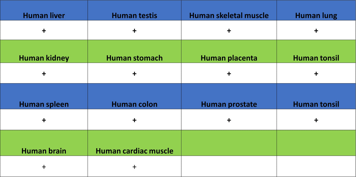
IHC shows positive staining in paraffin-embedded human kidney. Anti-CHKA antibody was used at 1/1000 dilution, followed by a HRP Polymer for Rabbit IgG (ready to use). Counterstained with hematoxylin. Heat mediated antigen retrieval with Tris/EDTA buffer pH9.0 was performed before commencing with IHC staining protocol.
IHC shows positive staining in paraffin-embedded human liver. Anti-CHKA antibody was used at 1/1000 dilution, followed by a HRP Polymer for Rabbit IgG (ready to use). Counterstained with hematoxylin. Heat mediated antigen retrieval with Tris/EDTA buffer pH9.0 was performed before commencing with IHC staining protocol.
IHC shows positive staining in paraffin-embedded human liver cancer. Anti-CHKA antibody was used at 1/1000 dilution, followed by a HRP Polymer for Rabbit IgG (ready to use). Counterstained with hematoxylin. Heat mediated antigen retrieval with Tris/EDTA buffer pH9.0 was performed before commencing with IHC staining protocol.