WB result of Chk2 Recombinant Rabbit mAb
Primary antibody: Chk2 Recombinant Rabbit mAb at 1/1000 dilution
Primary antibody incubation conditions: 1.5 hours at RT
Lane 1: HCT 116 whole cell lysate 25 μg
Lane 2: HeLa whole cell lysate 25 μg
Lane 3: HT-29 whole cell lysate 25 μg
Lane 4: RD whole cell lysate 25 μg
Lane 5: K-562 whole cell lysate 25 μg
Lane 6: Jurkat whole cell lysate 25 μg
Secondary antibody: Goat Anti-Rabbit IgG (H+L), HRP conjugated at 1/10000 dilution
Predicted MW: 61 kDa
Observed MW: 61 kDa
Chk2 Recombinant Rabbit mAb
Chk2 Recombinant Rabbit mAb
Price:
Regular price
$100 USD
Regular price
Sale price
$100 USD
Unit price
per
For shipping services or bulk orders, you may request a quotation.
Secure checkout with

View full details
Product Details
Product Details
Product Specification
| Host | Rabbit |
| Antigen | Chk2 |
| Location | Nucleus |
| Accession | O96017 |
| Antibody Type | Recombinant mAb |
| Isotype | IgG |
| Application | WB, IHC-P |
| Reactivity | Hu |
| Positive Sample | HCT 116, HeLa, HT-29, RD, K-562, Jurkat |
| Purification | Protein A |
| Concentration | 1 mg/ml |
| Conjugation | Unconjugated |
| Physical Appearance | Liquid |
| Storage Buffer | PBS, 40% Glycerol, 0.05% BSA, 0.02% sodium azide |
| Stability & Storage | 12 months from date of receipt / reconstitution, -20 °C as supplied |
Dilution
| application | dilution | species |
| WB | 1:1000 | Hu |
| IHC-P | 1:200 | Hu |
Background
Chk2 (checkpoint kinase 2) is a serine/threonine protein kinase encoded by the CHEK2 gene, containing 543 amino acids with a molecular weight of approximately 61 kDa, and is the human homolog of budding yeast Rad53p and fission yeast Cds1p. Its structure consists of three functional domains: an N-terminal SQ/TQ cluster domain (SCD), a central forkhead-associated domain (FHA), and a C-terminal kinase domain (KD). In cells without DNA damage, Chk2 exists as a monomer with its kinase activity in an inhibited state. When DNA double-strand breaks occur, the upstream kinase ATM phosphorylates Chk2 at Thr68, triggering Chk2 to form transient dimers through intermolecular interactions between phosphorylated Thr68 and the FHA domain. Dimerization further involves FHA-kinase domain and FHA-FHA interfaces, in which the Ile157 residue plays a central role in the FHA-kinase interface. Following dimer formation, the two kinase active sites are brought into close proximity, promoting trans-autophosphorylation of Thr383 in the activation loop and thereby fully activating kinase activity; the activated Chk2 then dissociates into monomers, and Thr68 phosphorylation is no longer required for maintaining activity. Activated Chk2 phosphorylates a variety of downstream substrates: it phosphorylates p53 at Ser20, releasing it from the inhibitor MDM2 and thereby transcriptionally activating cell cycle arrest genes such as p21 and initiating apoptosis; it phosphorylates Cdc25A at Ser123 to promote its degradation and inhibit S phase progression; it phosphorylates Cdc25C at Ser216 to block entry into mitosis; it also phosphorylates BRCA1 at Ser988 to promote its dissociation from Chk2 and participate in DNA repair, and induces apoptosis independently of p53 by phosphorylating PML. Germline mutations in CHEK2 are associated with a variant form of Li-Fraumeni syndrome and familial breast cancer susceptibility, among which the Nordic founder mutation 1100delC is the most common, and missense mutations such as Ile157Thr and Arg145Trp in the FHA domain have also been identified in cancer families.
Picture
Picture
Western Blot
Immunohistochemistry
IHC shows positive staining in paraffin-embedded human colon. Anti-Chk2 antibody was used at 1/200 dilution, followed by a HRP Polymer for Rabbit IgG (ready to use). Counterstained with hematoxylin. Heat mediated antigen retrieval with Tris/EDTA buffer pH9.0 was performed before commencing with IHC staining protocol.
IHC shows positive staining in paraffin-embedded human placenta. Anti-Chk2 antibody was used at 1/200 dilution, followed by a HRP Polymer for Rabbit IgG (ready to use). Counterstained with hematoxylin. Heat mediated antigen retrieval with Tris/EDTA buffer pH9.0 was performed before commencing with IHC staining protocol.
IHC shows positive staining in paraffin-embedded human testis. Anti-Chk2 antibody was used at 1/200 dilution, followed by a HRP Polymer for Rabbit IgG (ready to use). Counterstained with hematoxylin. Heat mediated antigen retrieval with Tris/EDTA buffer pH9.0 was performed before commencing with IHC staining protocol.
IHC shows positive staining in paraffin-embedded human tonsil. Anti-Chk2 antibody was used at 1/200 dilution, followed by a HRP Polymer for Rabbit IgG (ready to use). Counterstained with hematoxylin. Heat mediated antigen retrieval with Tris/EDTA buffer pH9.0 was performed before commencing with IHC staining protocol.
IHC shows positive staining in paraffin-embedded human transitional cell carcinoma. Anti-Chk2 antibody was used at 1/200 dilution, followed by a HRP Polymer for Rabbit IgG (ready to use). Counterstained with hematoxylin. Heat mediated antigen retrieval with Tris/EDTA buffer pH9.0 was performed before commencing with IHC staining protocol.
IHC shows positive staining in paraffin-embedded human cervical cancer. Anti-Chk2 antibody was used at 1/200 dilution, followed by a HRP Polymer for Rabbit IgG (ready to use). Counterstained with hematoxylin. Heat mediated antigen retrieval with Tris/EDTA buffer pH9.0 was performed before commencing with IHC staining protocol.
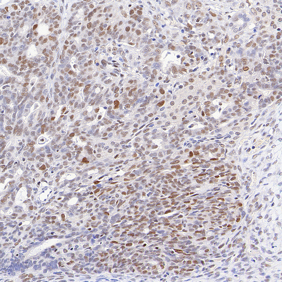
IHC shows positive staining in paraffin-embedded human ovarian cancer. Anti-Chk2 antibody was used at 1/200 dilution, followed by a HRP Polymer for Rabbit IgG (ready to use). Counterstained with hematoxylin. Heat mediated antigen retrieval with Tris/EDTA buffer pH9.0 was performed before commencing with IHC staining protocol.