WB result of Chk1 Recombinant Mouse mAb
Primary antibody: Chk1 Recombinant Mouse mAb at 1/1000 dilution
Primary antibody incubation conditions: overnight at 4°C
Lane 1: SK-MEL-28 whole cell lysate 20 μg
Lane 2: 293T whole cell lysate 20 μg
Lane 3: HeLa whole cell lysate 20 μg
Lane 4: A431 whole cell lysate 20 μg
Secondary antibody: Goat Anti-Mouse IgG (H+L), HRP conjugated at 1/10000 dilution
Predicted MW: 54 kDa
Observed MW: 54 kDa
This blot was developed with high sensitivity substrate
Chk1 Recombinant Mouse mAb (S-6263)
Chk1 Recombinant Mouse mAb (S-6263)
Price:
Regular price
$100 USD
Regular price
Sale price
$100 USD
Unit price
per
For shipping services or bulk orders, you may request a quotation.
Secure checkout with

View full details
Product Details
Product Details
Product Specification
| Host | Mouse |
| Antigen | Chk1 |
| Location | Cytoplasm, Cytoskeleton, Nucleus |
| Accession | O14757 |
| Clone Number | S-6263 |
| Antibody Type | Recombinant mAb |
| Isotype | IgG1 |
| Application | WB, IHC-P |
| Reactivity | Hu, Ms, Rt |
| Positive Sample | SK-MEL-28, HeLa, 293T, A431, NIH/3T3 |
| Predicted Reactivity | Mk |
| Purification | Protein A |
| Concentration | 2 mg/ml |
| Conjugation | Unconjugated |
| Physical Appearance | Liquid |
| Storage Buffer | PBS, 40% Glycerol, 0.05% BSA, 0.02% sodium azide |
| Stability & Storage | 12 months from date of receipt / reconstitution, -20 °C as supplied |
Dilution
| application | dilution | species |
| WB | 1:1000 | Hu, Ms |
| IHC-P | 1:500 | Hu, Ms, Rt |
Background
Chk1 (checkpoint kinase 1) is a serine/threonine protein kinase encoded by the CHEK1 gene located on chromosome 11q24, with a molecular weight of approximately 54 kDa and 476 amino acids. It is highly conserved in evolution, with homologous proteins present from fission yeast to humans. As a core effector molecule in the DNA damage response pathway, Chk1 is a direct substrate of the upstream sensor kinase ATR and is activated by ATR phosphorylation upon DNA replication stress or damage, thereby transmitting checkpoint signals by phosphorylating downstream target proteins such as the CDC25 phosphatase family. The primary function of Chk1 is to exert regulatory control during the S phase and G2/M phase of the cell cycle, delaying mitotic entry by inhibiting CDK activity to provide a time window for DNA repair, thereby maintaining genomic stability. In addition, Chk1 participates in regulating centrosome and spindle checkpoints, inhibiting the activation of DNA replication origins, and suppressing the transcription of cell cycle proteins such as cyclin B and CDK1. In malignant tumors, Chk1 is often abnormally overexpressed, enhancing the DNA repair capacity of tumor cells and inducing resistance to chemotherapy and radiotherapy, making it an important molecular target for cancer therapy. Several Chk1 inhibitors have entered clinical trials, among which Prexasertib has been granted Fast Track designation by the FDA for the treatment of patients with platinum-resistant ovarian cancer and endometrial cancer.
Picture
Picture
Western Blot
WB result of Chk1 Recombinant Mouse mAb
Primary antibody: Chk1 Recombinant Mouse mAb at 1/1000 dilution
Primary antibody incubation conditions: overnight at 4°C
Lane 1: NIH/3T3 whole cell lysate 20 μg
Secondary antibody: Goat Anti-Mouse IgG (H+L), HRP conjugated at 1/10000 dilution
Predicted MW: 54 kDa
Observed MW: 54 kDa
This blot was developed with high sensitivity substrate
Immunohistochemistry
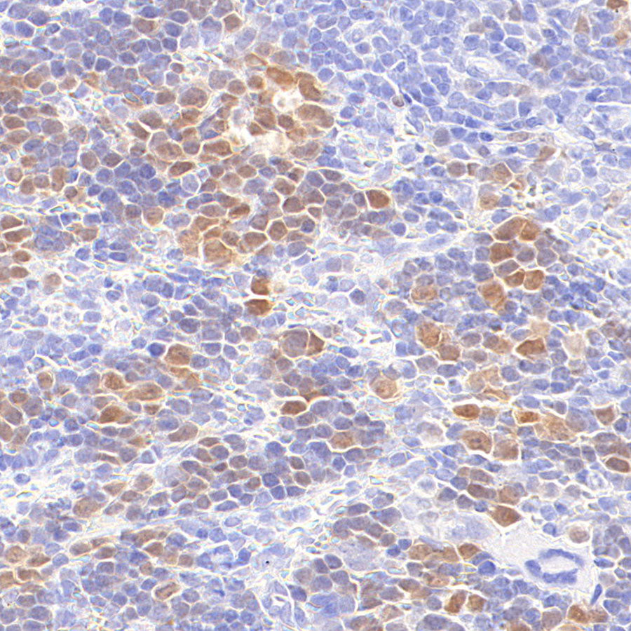
IHC shows positive staining in paraffin-embedded human tonsil. Anti-Chk1 antibody was used at 1/500 dilution, followed by a HRP Polymer for Mouse IgG (ready to use). Counterstained with hematoxylin. Heat mediated antigen retrieval with Tris/EDTA buffer pH9.0 was performed before commencing with IHC staining protocol.
IHC shows positive staining in paraffin-embedded human thymoma. Anti-Chk1 antibody was used at 1/500 dilution, followed by a HRP Polymer for Mouse IgG (ready to use). Counterstained with hematoxylin. Heat mediated antigen retrieval with Tris/EDTA buffer pH9.0 was performed before commencing with IHC staining protocol.
IHC shows positive staining in paraffin-embedded mouse spleen. Anti-Chk1 antibody was used at 1/500 dilution, followed by a HRP Polymer for Mouse IgG (ready to use). Counterstained with hematoxylin. Heat mediated antigen retrieval with Tris/EDTA buffer pH9.0 was performed before commencing with IHC staining protocol.
IHC shows positive staining in paraffin-embedded rat spleen. Anti-Chk1 antibody was used at 1/500 dilution, followed by a HRP Polymer for Mouse IgG (ready to use). Counterstained with hematoxylin. Heat mediated antigen retrieval with Tris/EDTA buffer pH9.0 was performed before commencing with IHC staining protocol.