WB result of CEBPA Recombinant Rabbit mAb
Primary antibody: CEBPA Recombinant Rabbit mAb at 1/1000 dilution
Lane 1: Jurkat whole cell lysate 20 µg
Lane 2: HepG2 whole cell lysate 20 µg
Lane 3: LNCaP whole cell lysate 20 µg
Lane 4: THP-1 whole cell lysate 20 µg
Lane 5: HL-60 whole cell lysate 20 µg
Low expression control: Jurkat whole cell lysate
Secondary antibody: Goat Anti- rabbit IgG, (H+L), HRP conjugated at 1/10000 dilution
Predicted MW: 37 kDa
Observed MW: 26, 34, 40 kDa
CEBPA Recombinant Rabbit mAb (S-3629-33)
CEBPA Recombinant Rabbit mAb (S-3629-33)
Price:
Regular price
$100 USD
Regular price
Sale price
$100 USD
Unit price
per
For shipping services or bulk orders, you may request a quotation.
Secure checkout with

View full details
Product Details
Product Details
Product Specification
| Host | Rabbit |
| Antigen | CEBPA |
| Synonyms | CCAAT/enhancer-binding protein alpha; CEBP |
| Immunogen | Recombinant Protein |
| Location | Nucleus |
| Accession | P49715 |
| Clone Number | S-3629-33 |
| Antibody Type | Recombinant mAb |
| Isotype | IgG |
| Application | WB, IHC-P, ICC |
| Reactivity | Hu, Ms, Rt |
| Positive Sample | HepG2, LnCaP, THP-1, HL-60 |
| Purification | Protein A |
| Concentration | 0.5 mg/ml |
| Conjugation | Unconjugated |
| Physical Appearance | Liquid |
| Storage Buffer | PBS, 40% Glycerol, 0.05% BSA, 0.03% Proclin 300 |
| Stability & Storage | 12 months from date of receipt / reconstitution, -20 °C as supplied |
Dilution
| application | dilution | species |
| WB | 1:1000 | Hu |
| IHC-P | 1:500 | Hu, Ms, Rt |
| ICC | 1:500 | Hu |
Background
CEBPA protein (CCAAT/enhancer-binding protein alpha) is a transcription factor that plays a central role in regulating cell differentiation and proliferation, and is an important member of the basic leucine zipper transcription factor family. This protein is primarily highly expressed in organs such as the liver, adipose tissue, bone marrow, and lungs. By binding to specific DNA sequences in the promoter regions of target genes, it activates or represses the expression of various genes involved in cell cycle arrest, terminal differentiation, and metabolic regulation. In the hematopoietic system, CEBPA is a key regulator of granulocytic differentiation, promoting the differentiation of myeloid progenitors into mature blood cells such as neutrophils; in the liver, it participates in regulating glucose and lipid metabolism as well as the expression of acute-phase response proteins; in adipogenesis, it cooperates with other transcription factors to drive preadipocyte differentiation into mature adipocytes. Furthermore, CEBPA directly blocks cell proliferation by inhibiting cyclins (such as through the E2F pathway). Loss-of-function mutations leading to CEBPA inactivation are closely associated with the development of malignant tumors such as acute myeloid leukemia (AML)—the so-called "CEBPA double mutation" has been considered a favorable prognostic marker in certain types of AML. Abnormal expression of this protein is also linked to metabolic diseases, liver fibrosis, and related conditions. Therefore, CEBPA is not only a key guardian of normal tissue differentiation homeostasis but also a hot topic target in tumorigenesis and regenerative medicine research.
Picture
Picture
Western Blot
Immunohistochemistry
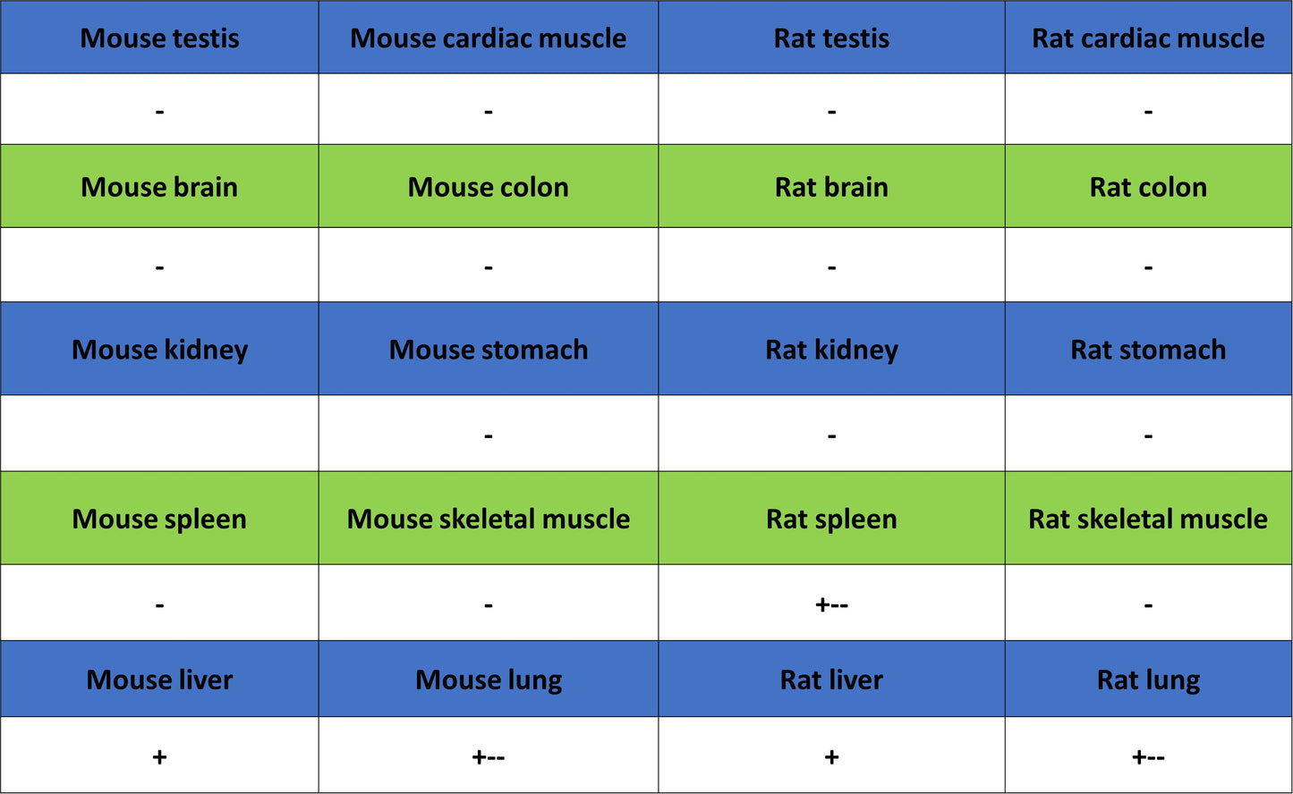
IHC shows positive staining in paraffin-embedded human endometrial cancer. Anti-CEBPA antibody was used at 1/500 dilution, followed by a HRP Polymer for Rabbit IgG (ready to use). Counterstained with hematoxylin. Heat mediated antigen retrieval with Tris/EDTA buffer pH9.0 was performed before commencing with IHC staining protocol.
IHC shows positive staining in paraffin-embedded human spleen. Anti-CEBPA antibody was used at 1/500 dilution, followed by a HRP Polymer for Rabbit IgG (ready to use). Counterstained with hematoxylin. Heat mediated antigen retrieval with Tris/EDTA buffer pH9.0 was performed before commencing with IHC staining protocol.
IHC shows positive staining in paraffin-embedded human tonsil. Anti-CEBPA antibody was used at 1/500 dilution, followed by a HRP Polymer for Rabbit IgG (ready to use). Counterstained with hematoxylin. Heat mediated antigen retrieval with Tris/EDTA buffer pH9.0 was performed before commencing with IHC staining protocol.
IHC shows positive staining in paraffin-embedded human placenta. Anti-CEBPA antibody was used at 1/500 dilution, followed by a HRP Polymer for Rabbit IgG (ready to use). Counterstained with hematoxylin. Heat mediated antigen retrieval with Tris/EDTA buffer pH9.0 was performed before commencing with IHC staining protocol.
IHC shows positive staining in paraffin-embedded mouse liver. Anti-CEBPA antibody was used at 1/500 dilution, followed by a HRP Polymer for Rabbit IgG (ready to use). Counterstained with hematoxylin. Heat mediated antigen retrieval with Tris/EDTA buffer pH9.0 was performed before commencing with IHC staining protocol.
IHC shows positive staining in paraffin-embedded rat spleen. Anti-CEBPA antibody was used at 1/500 dilution, followed by a HRP Polymer for Rabbit IgG (ready to use). Counterstained with hematoxylin. Heat mediated antigen retrieval with Tris/EDTA buffer pH9.0 was performed before commencing with IHC staining protocol.
Immunocytochemistry
ICC shows positive staining in THP-1 cells (top panel) and negative staining in Jurkat cells (below panel). Anti-CEBPA antibody was used at 1/500 dilution (Green) and incubated overnight at 4°C. Goat polyclonal Antibody to Rabbit IgG - H&L (Alexa Fluor® 488) was used as secondary antibody at 1/1000 dilution. The cells were fixed with 4% PFA and permeabilized with 0.1% PBS-Triton X-100. Nuclei were counterstained with DAPI (Blue). Counterstain with tubulin (Red).