WB result of CEA Recombinant Rabbit mAb
Primary antibody: CEA Recombinant Rabbit mAb at 1/1000 dilution
Lane 1: PANC-1 whole cell lysate 20 µg
Lane 2: MCF7 whole cell lysate 20 µg
Lane 3: HT-29 whole cell lysate 20 µg
Lane 4: BxPC-3 whole cell lysate 20 µg
Negative control: PANC-1 whole cell lysate
Secondary antibody: Goat Anti- rabbit IgG, (H+L), HRP conjugated at 1/10000 dilution
Predicted MW: 77 kDa
Observed MW: 150-300 kDa
CEA/CEACAM5 Recombinant Rabbit mAb (SDT-098-41)
CEA/CEACAM5 Recombinant Rabbit mAb (SDT-098-41)
Price:
Regular price
$100 USD
Regular price
Sale price
$100 USD
Unit price
per
For shipping services or bulk orders, you may request a quotation.
Secure checkout with

View full details
Product Details
Product Details
Product Specification
| Host | Rabbit |
| Antigen | CEA |
| Synonyms | CEACAM5,Cell adhesion molecule CEACAM5; Carcinoembryonic antigen (CEA); Carcinoembryonic antigen-related cell adhesion molecule 5 (CEA cell adhesion molecule 5Imported); Meconium antigen 100; CD66e; CEA s; CEACAM5 |
| Immunogen | Recombinant Protein |
| Location | Cell membrane |
| Accession | P06731 |
| Clone Number | SDT-098-41 |
| Antibody Type | Recombinant mAb |
| Isotype | IgG |
| Application | WB, IHC-P |
| Reactivity | Hu |
| Positive Sample | MCF7, HT-29, BxPC-3 |
| Purification | Protein A |
| Concentration | 0.1 mg/ml |
| Conjugation | Unconjugated |
| Physical Appearance | Liquid |
| Storage Buffer | PBS, 40% Glycerol, 0.05% BSA, 0.02% sodium azide |
| Stability & Storage | 12 months from date of receipt / reconstitution, -20 °C as supplied |
Dilution
| application | dilution | species |
| WB | 1:500-1:1000 | Hu |
| IHC-P | 1:1000-1:2000 | Hu |
Background
CEACAM5 (Carcinoembryonic Antigen-Related Cell Adhesion Molecule 5), commonly known as CEA (Carcinoembryonic Antigen), is an acidic glycoprotein widely expressed in human epithelial tissues and belongs to the immunoglobulin superfamily. Structurally, it is anchored to the cell membrane via a glycosylphosphatidylinositol (GPI) moiety and lacks an intracellular signal transduction domain. Under physiological conditions, CEACAM5 is highly expressed only in the gastrointestinal tract, pancreas, and liver during early fetal development, with expression levels significantly decreasing after birth. However, under pathological conditions, particularly in various malignant tumors such as colorectal cancer, gastric cancer, lung cancer, pancreatic cancer, and breast cancer, CEACAM5 is frequently overexpressed. As a classic tumor marker, serum CEACAM5 levels have been widely used for cancer clinical diagnosis, treatment efficacy monitoring, and recurrence prediction. Functionally, as a cell adhesion molecule, CEACAM5 primarily regulates tumor cell aggregation, polarity, migration, and invasion through homophilic or heterophilic interactions, while also inhibiting differentiation and blocking anoikis, thereby promoting tumor metastasis and drug resistance. Due to its highly specific expression in tumor tissues, CEACAM5 has become an important target for antibody-based targeted therapy and immunotherapy.
Picture
Picture
Western Blot
Immunohistochemistry
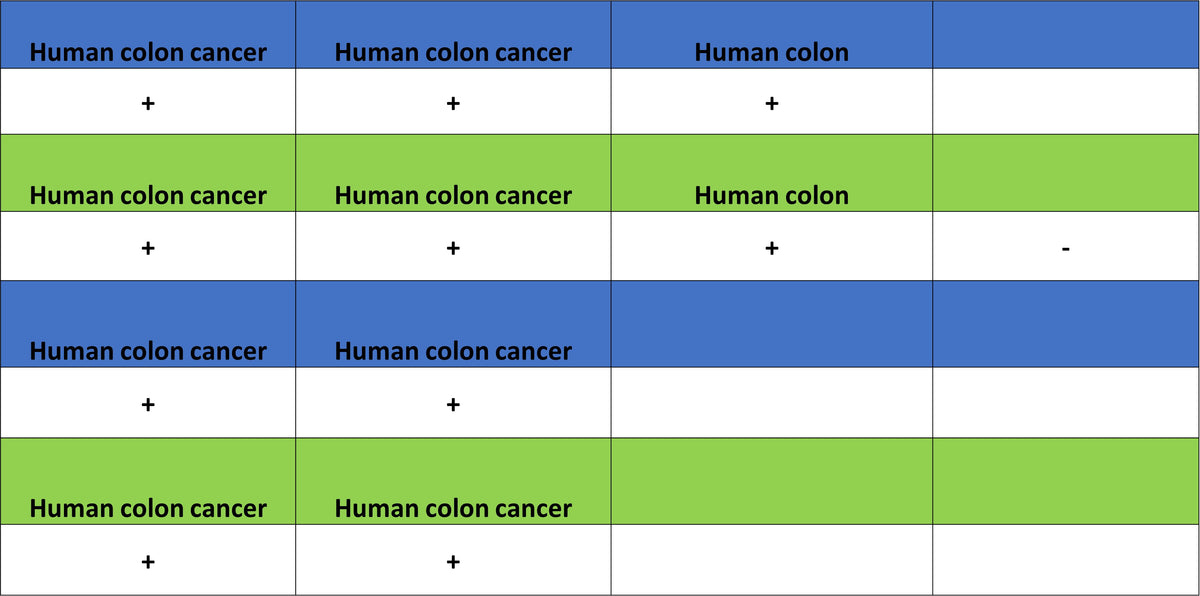
IHC shows positive staining in paraffin-embedded human colon. Anti-CEA antibody was used at 1/1200 dilution, followed by a HRP Polymer for Rabbit IgG (ready to use). Counterstained with hematoxylin. Heat mediated antigen retrieval with Tris/EDTA buffer pH9.0 was performed before commencing with IHC staining protocol.
IHC shows positive staining in paraffin-embedded human colon cancer. Anti-CEA antibody was used at 1/1200 dilution, followed by a HRP Polymer for Rabbit IgG (ready to use). Counterstained with hematoxylin. Heat mediated antigen retrieval with Tris/EDTA buffer pH9.0 was performed before commencing with IHC staining protocol.
IHC shows positive staining in paraffin-embedded human lung adenocarcinoma. Anti-CEA antibody was used at 1/1200 dilution, followed by a HRP Polymer for Rabbit IgG (ready to use). Counterstained with hematoxylin. Heat mediated antigen retrieval with Tris/EDTA buffer pH9.0 was performed before commencing with IHC staining protocol.
IHC shows positive staining in paraffin-embedded human pancreatic cancer. Anti-CEA antibody was used at 1/1200 dilution, followed by a HRP Polymer for Rabbit IgG (ready to use). Counterstained with hematoxylin. Heat mediated antigen retrieval with Tris/EDTA buffer pH9.0 was performed before commencing with IHC staining protocol.