WB result of CDK11/CDC2L2/CDK11A Recombinant Rabbit mAb
Primary antibody: CDK11/CDC2L2/CDK11A Recombinant Rabbit mAb at 1/1000 dilution
Lane 1: HeLa whole cell lysate 5 µg
Lane 2: 293T whole cell lysate 5 µg
Lane 3: Raji whole cell lysate 5 µg
Lane 4: HepG2 whole cell lysate 5 µg
Lane 5: A431 whole cell lysate 5 µg
Lane 6: A375 whole cell lysate 5 µg
Lane 7: MCF7 whole cell lysate 5 µg
Lane 8: MDA-MB-231 whole cell lysate 5 µg
Secondary antibody: Goat Anti- rabbit IgG, (H+L), HRP conjugated at 1/10000 dilution
Predicted MW: 91 kDa
Observed MW: 120 kDa
CDK11/CDC2L2/CDK11A Recombinant Rabbit mAb (S-3994-75)
CDK11/CDC2L2/CDK11A Recombinant Rabbit mAb (S-3994-75)
Price:
Regular price
$100 USD
Regular price
Sale price
$100 USD
Unit price
per
For shipping services or bulk orders, you may request a quotation.
Secure checkout with

View full details
Product Details
Product Details
Product Specification
| Host | Rabbit |
| Antigen | CDK11/CDC2L2/CDK11A |
| Synonyms | Cyclin-dependent kinase 11A; Cell division cycle 2-like protein kinase 2; Cell division protein kinase 11A; Galactosyltransferase-associated protein kinase p58/GTA; PITSLRE serine/threonine-protein kinase CDC2L2; CDC2L2; CDC2L3; PITSLREB |
| Immunogen | Recombinant Protein |
| Location | Cytoplasm, Nucleus |
| Accession | Q9UQ88 |
| Clone Number | S-3994-75 |
| Antibody Type | Recombinant mAb |
| Isotype | IgG |
| Application | WB, IHC-P |
| Reactivity | Hu, Ms, Rt |
| Positive Sample | HeLa, 293T, Raji, HepG2, A431, A375, MCF7, MDA-MB-231, NIH/3T3, C2C12, C6, rat testis |
| Purification | Protein A |
| Concentration | 0.5 mg/ml |
| Conjugation | Unconjugated |
| Physical Appearance | Liquid |
| Storage Buffer | PBS, 40% Glycerol, 0.05% BSA, 0.02% sodium azide |
| Stability & Storage | 12 months from date of receipt / reconstitution, -20 °C as supplied |
Dilution
| application | dilution | species |
| WB | 1:1000-1:2000 | Hu, Ms, Rt |
| IHC-P | 1:500 | Hu, Ms, Rt |
Background
CDK11 (Cyclin-Dependent Kinase 11) is a serine/threonine protein kinase that plays a core role in the regulation of transcription and pre-mRNA splicing, and is a member of the CDK family that is relatively less studied but essential for the survival of metazoans. In human cells, CDK11 is encoded by two nearly identical (97.5% homologous) genes, CDK11A and CDK11B, which produce three major functional isoforms through alternative splicing and translational regulation, each undertaking distinct cellular functions. The full-length isoform CDK11p110 is continuously expressed throughout the cell cycle and is primarily localized in the nucleus, where it binds to Cyclin L, phosphorylates the spliceosome core component SF3B1 to regulate spliceosome activation and constitutive splicing, and also participates in the transcriptional regulation of specific genes (such as replication-dependent histone genes) through phosphorylation of the C-terminal domain (CTD) of RNA polymerase II. The CDK11p58 isoform is transiently expressed only during the G2/M phase of the cell cycle; upon binding to Cyclin D3, it localizes to centrosomes and is responsible for centrosome maturation and bipolar spindle assembly during mitosis, which is critical for maintaining sister chromatid cohesion. The CDK11p46 isoform, on the other hand, is an apoptosis-specific product generated by caspase-mediated cleavage of the precursor isoform, primarily localized in the cytoplasm, and participates in regulating the apoptotic process by phosphorylating targets such as translation initiation factors. Given the critical role of CDK11 in gene expression and cell proliferation, its dysfunction is closely associated with various cancers. The selective CDK11 inhibitor OTS964 has emerged as a highly promising anti-tumor targeted therapeutic strategy, showing favorable prospects in preclinical studies.
Picture
Picture
Western Blot
WB result of CDK11/CDC2L2/CDK11A Recombinant Rabbit mAb
Primary antibody: CDK11/CDC2L2/CDK11A Recombinant Rabbit mAb at 1/1000 dilution
Lane 1: NIH/3T3 whole cell lysate 5 µg
Lane 2: C2C12 whole cell lysate 5 µg
Secondary antibody: Goat Anti- rabbit IgG, (H+L), HRP conjugated at 1/10000 dilution
Predicted MW: 91 kDa
Observed MW: 120 kDa
WB result of CDK11/CDC2L2/CDK11A Recombinant Rabbit mAb
Primary antibody: CDK11/CDC2L2/CDK11A Recombinant Rabbit mAb at 1/1000 dilution
Lane 1: C6 whole cell lysate 5 µg
Lane 2: rat testis lysate 5 µg
Secondary antibody: Goat Anti- rabbit IgG, (H+L), HRP conjugated at 1/10000 dilution
Predicted MW: 91 kDa
Observed MW: 120 kDa
Immunohistochemistry
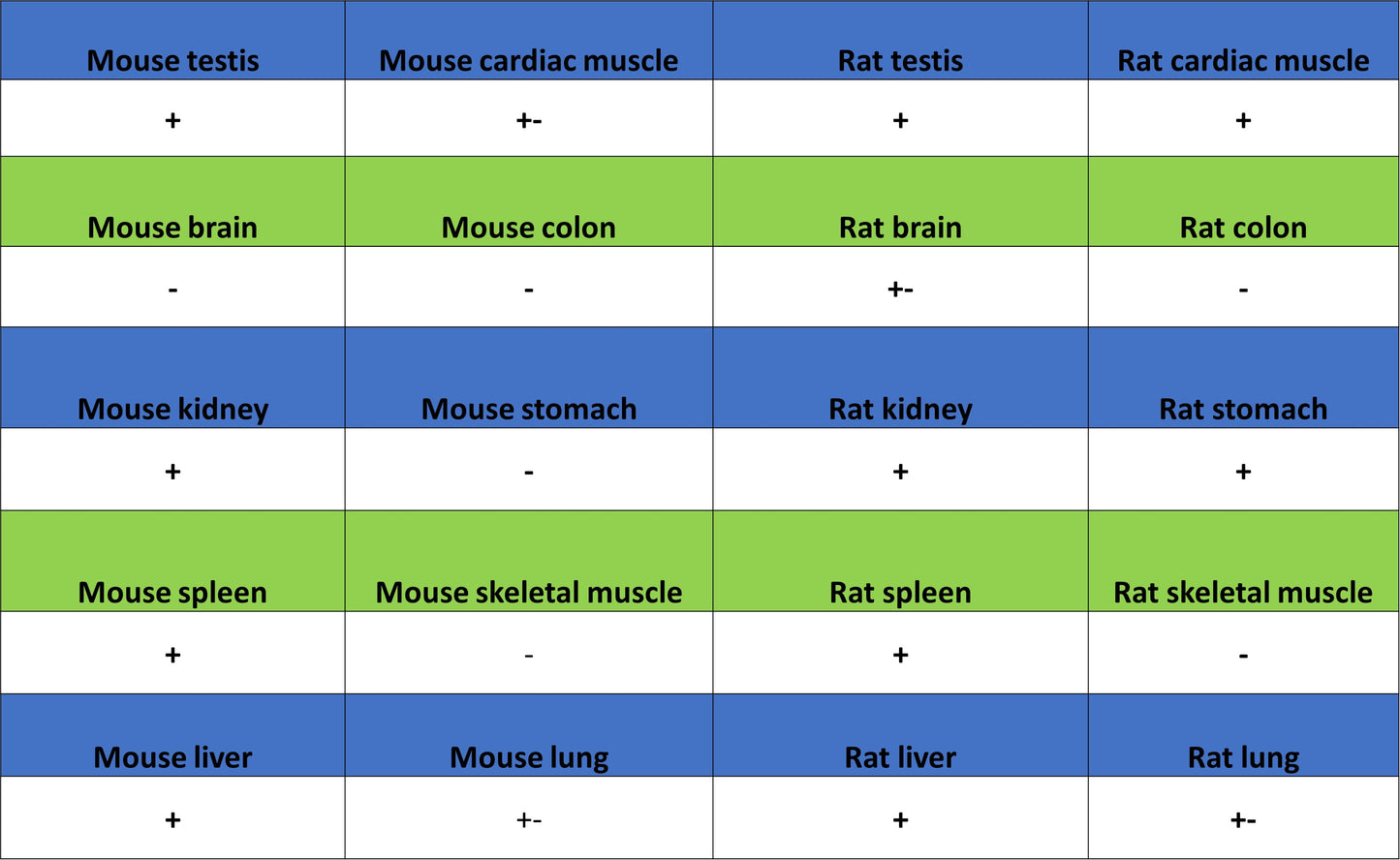
IHC shows positive staining in paraffin-embedded human colon. Anti-CDK11 antibody was used at 1/500 dilution, followed by a HRP Polymer for Rabbit IgG (ready to use). Counterstained with hematoxylin. Heat mediated antigen retrieval with Tris/EDTA buffer pH9.0 was performed before commencing with IHC staining protocol.
IHC shows positive staining in paraffin-embedded human spleen. Anti-CDK11 antibody was used at 1/500 dilution, followed by a HRP Polymer for Rabbit IgG (ready to use). Counterstained with hematoxylin. Heat mediated antigen retrieval with Tris/EDTA buffer pH9.0 was performed before commencing with IHC staining protocol.
IHC shows positive staining in paraffin-embedded human lung cancer. Anti-CDK11 antibody was used at 1/500 dilution, followed by a HRP Polymer for Rabbit IgG (ready to use). Counterstained with hematoxylin. Heat mediated antigen retrieval with Tris/EDTA buffer pH9.0 was performed before commencing with IHC staining protocol.
IHC shows positive staining in paraffin-embedded mouse testis. Anti-CDK11 antibody was used at 1/500 dilution, followed by a HRP Polymer for Rabbit IgG (ready to use). Counterstained with hematoxylin. Heat mediated antigen retrieval with Tris/EDTA buffer pH9.0 was performed before commencing with IHC staining protocol.
IHC shows positive staining in paraffin-embedded rat testis. Anti-CDK11 antibody was used at 1/500 dilution, followed by a HRP Polymer for Rabbit IgG (ready to use). Counterstained with hematoxylin. Heat mediated antigen retrieval with Tris/EDTA buffer pH9.0 was performed before commencing with IHC staining protocol.