WB result of CDH3 Recombinant Rabbit mAb
Primary antibody: CDH3 Recombinant Rabbit mAb at 1/1000 dilution
Lane 1: MCF7 whole cell lysate 20 µg
Lane 2: A431 whole cell lysate 20 µg
Lane 3: BxPC-3 whole cell lysate 20 µg
Lane 4: HCT 116 whole cell lysate 20 µg
Negative control: MCF7 whole cell lysate
Secondary antibody: Goat Anti- rabbit IgG, (H+L), HRP conjugated at 1/10000 dilution
Predicted MW: 91 kDa
Observed MW: 125 kDa
CDH3 Recombinant Rabbit mAb (S-3576-116)
CDH3 Recombinant Rabbit mAb (S-3576-116)
Price:
Regular price
$100 USD
Regular price
Sale price
$100 USD
Unit price
per
For shipping services or bulk orders, you may request a quotation.
Secure checkout with

View full details
Product Details
Product Details
Product Specification
| Host | Rabbit |
| Antigen | CDH3 |
| Synonyms | Cadherin-3; CDHP |
| Immunogen | Synthetic Peptide |
| Location | Cell membrane |
| Accession | P22223 |
| Clone Number | S-3576-116 |
| Antibody Type | Recombinant mAb |
| Isotype | IgG |
| Application | WB, IHC-P |
| Reactivity | Hu, Ms |
| Purification | Protein A |
| Concentration | 0.5 mg/ml |
| Conjugation | Unconjugated |
| Physical Appearance | Liquid |
| Storage Buffer | PBS, 40% Glycerol, 0.05% BSA, 0.02% sodium azide |
| Stability & Storage | 12 months from date of receipt / reconstitution, -20 °C as supplied |
Dilution
| application | dilution | species |
| WB | 1:1000 | Hu, Ms |
| IHC-P | 1:500 | Hu |
Background
CDH3, also known as P-cadherin (Placental cadherin), is a classical cadherin family member encoded by the CDH3 gene, functioning as a calcium-dependent, single-pass transmembrane cell adhesion glycoprotein. Its precursor protein consists of 829 amino acids and is processed by proteolytic cleavage to form a mature protein of approximately 118 kDa. Structurally, it comprises five extracellular cadherin repeats (with the N-terminal EC1 domain being particularly critical for adhesion specificity) responsible for calcium-dependent homophilic adhesion, a transmembrane region, and a highly conserved intracellular domain that links to the actin cytoskeleton through interactions with β-catenin and other proteins. Under physiological conditions, CDH3 expression exhibits high spatiotemporal specificity, being predominantly expressed in the basal layer proliferative cells of certain epithelial tissues, and plays a critical role in embryonic development, hair follicle cycling, and maintenance of normal mammary gland structure. Functional abnormalities or gene mutations in CDH3 can lead to hereditary disorders such as hypotrichosis with juvenile macular dystrophy or ectodermal dysplasia-syndactyly-macular dystrophy syndrome. In tumor biology, the function of CDH3 exhibits significant context-dependent bidirectionality: on the one hand, in various malignancies including breast cancer, gastric cancer, and pancreatic cancer, CDH3 is often aberrantly overexpressed, driving malignant progression by promoting cell migration, invasion, and epithelial–mesenchymal transition, and is closely associated with poor patient prognosis; on the other hand, in certain cancer types or specific microenvironments, it may also play a tumor-suppressive role. Based on its tumor-specific expression, therapeutic strategies targeting CDH3, including monoclonal antibodies and antibody-drug conjugates, have demonstrated promising potential in preclinical and early-stage clinical trials.
Picture
Picture
Western Blot
WB result of CDH3 Recombinant Rabbit mAb
Primary antibody: CDH3 Recombinant Rabbit mAb at 1/1000 dilution
Lane 1: mouse placenta lysate 20 µg
Secondary antibody: Goat Anti- rabbit IgG, (H+L), HRP conjugated at 1/10000 dilution
Predicted MW: 91 kDa
Observed MW: 125 kDa
Immunohistochemistry
IHC shows positive staining in paraffin-embedded human breast. Anti-CDH3 antibody was used at 1/500 dilution, followed by a HRP Polymer for Rabbit IgG (ready to use). Counterstained with hematoxylin. Heat mediated antigen retrieval with Tris/EDTA buffer pH9.0 was performed before commencing with IHC staining protocol.
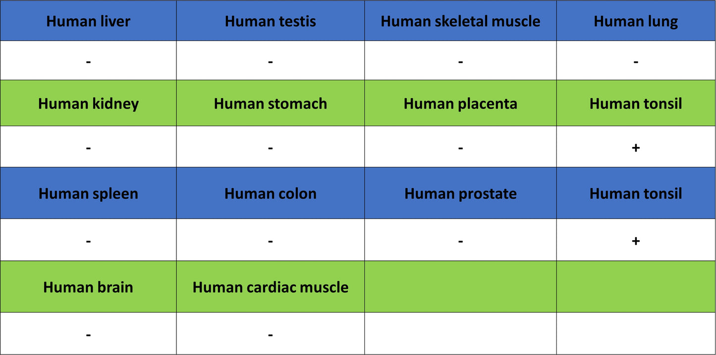
IHC shows positive staining in paraffin-embedded human tonsil. Anti-CDH3 antibody was used at 1/500 dilution, followed by a HRP Polymer for Rabbit IgG (ready to use). Counterstained with hematoxylin. Heat mediated antigen retrieval with Tris/EDTA buffer pH9.0 was performed before commencing with IHC staining protocol.
IHC shows positive staining in paraffin-embedded human cervical squamous cell carcinoma. Anti-CDH3 antibody was used at 1/500 dilution, followed by a HRP Polymer for Rabbit IgG (ready to use). Counterstained with hematoxylin. Heat mediated antigen retrieval with Tris/EDTA buffer pH9.0 was performed before commencing with IHC staining protocol.
IHC shows positive staining in paraffin-embedded human colon cancer. Anti-CDH3 antibody was used at 1/500 dilution, followed by a HRP Polymer for Rabbit IgG (ready to use). Counterstained with hematoxylin. Heat mediated antigen retrieval with Tris/EDTA buffer pH9.0 was performed before commencing with IHC staining protocol.