CDCP1 Recombinant Rabbit mAb (S-3437-221)
CDCP1 Recombinant Rabbit mAb (S-3437-221)
Price:
Regular price
$100 USD
Regular price
Sale price
$100 USD
Unit price
per
For shipping services or bulk orders, you may request a quotation.
Secure checkout with

View full details
Product Details
Product Details
Product Specification
| Host | Rabbit |
| Antigen | CDCP1 |
| Synonyms | CUB domain-containing protein 1; Membrane glycoprotein gp140; Subtractive immunization M plus HEp3-associated 135 kDa protein (SIMA135); Transmembrane and associated with src kinases; CD318; TRASK |
| Immunogen | Recombinant Protein |
| Location | Secreted, Cell membrane |
| Accession | Q9H5V8 |
| Clone Number | S-3437-221 |
| Antibody Type | Recombinant mAb |
| Isotype | IgG |
| Application | WB, IHC-P |
| Reactivity | Hu |
| Positive Sample | PC-3, HCT 116, PANC-1, A549 |
| Purification | Protein A |
| Concentration | 0.5 mg/ml |
| Conjugation | Unconjugated |
| Physical Appearance | Liquid |
| Storage Buffer | PBS, 40% Glycerol, 0.05% BSA, 0.02% sodium azide |
| Stability & Storage | 12 months from date of receipt / reconstitution, -20 °C as supplied |
Dilution
| application | dilution | species |
| WB | 1:500-1:1000 | Hu |
| IHC-P | 1:2000 | Hu |
Background
CDCP1 (CUB domain-containing protein 1), also known as CD318 or SIMA135, is a type I single-pass transmembrane glycoprotein characterized by three CUB domains in its extracellular region and tyrosine phosphorylation sites in its intracellular domain that interact with Src family kinases. In normal tissues, it is primarily expressed in skeletal muscle, colon, and certain stem or progenitor cell populations, but its most prominent function has been observed in multiple human cancers. As a substrate for signaling molecules such as Src kinases, CDCP1, upon tyrosine phosphorylation, initiates downstream signaling pathways that promote tumor cell migration, invasion, anoikis resistance, and metastasis. Recent studies have revealed more complex mechanisms, such as its role as a novel co-receptor for TGF-β1, enhancing the pro-tumorigenic TGF-β/Smad signaling pathway; it has also been found to drive malignant tumor progression through activation of signaling axes such as Wnt/β-catenin. Its high expression in various solid tumors, including lung, colorectal, and breast cancers, is closely associated with poor patient prognosis, making it a highly promising anti-tumor therapeutic target and cancer diagnostic biomarker.
Picture
Picture
Western Blot
WB result of CDCP1 Recombinant Rabbit mAb
Primary antibody: CDCP1 Recombinant Rabbit mAb Rabbit mAb at 1/1000 dilution
Lane 1: MCF7 whole cell lysate 20 µg
Lane 2: PC-3 whole cell lysate 20 µg
Lane 3: HCT 116 whole cell lysate 20 µg
Lane 4: PANC-1 whole cell lysate 20 µg
Lane 5: A431 whole cell lysate 20 µg
Negative control: MCF7 whole cell lysate
Secondary antibody: Goat Anti-Rabbit IgG, (H+L), HRP conjugated at 1/10000 dilution
Predicted MW: 93 kDa
Observed MW: 70, 140 kDa
This blot was developed with high sensitivity substrate
Immunohistochemistry
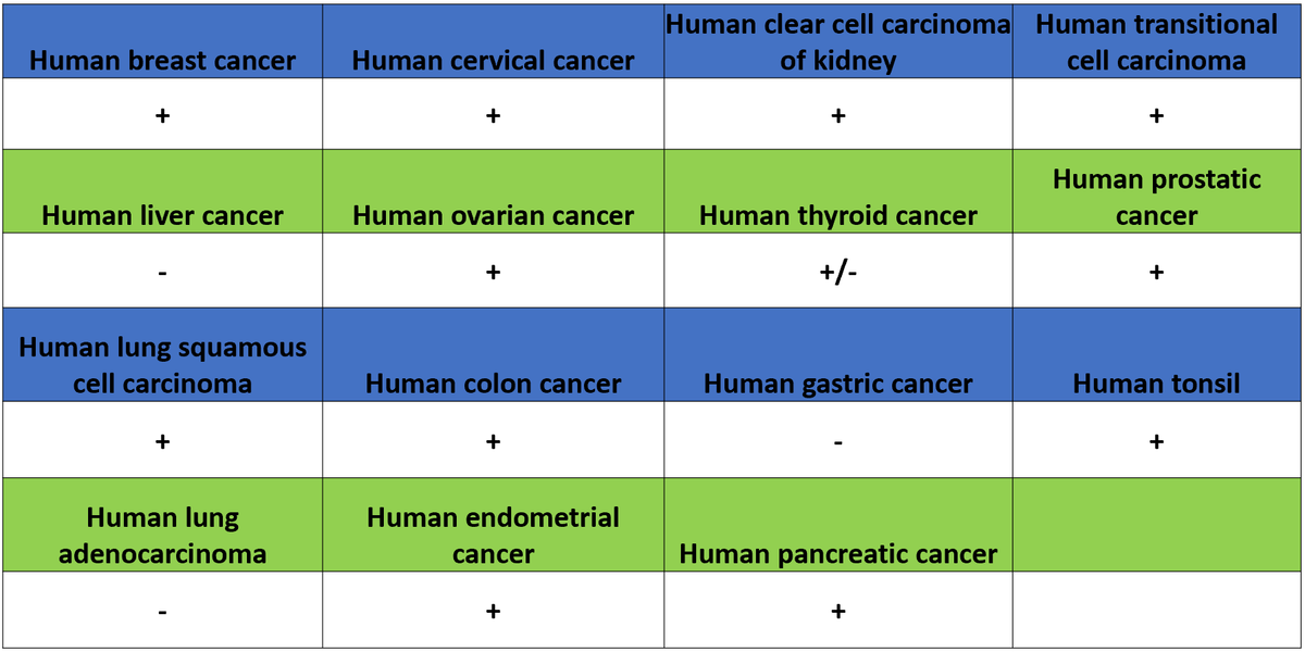
IHC shows positive staining in paraffin-embedded human colon cancer. Anti-CDCP1 antibody was used at 1/2000 dilution, followed by a HRP Polymer for Rabbit IgG (ready to use). Counterstained with hematoxylin. Heat mediated antigen retrieval with Tris/EDTA buffer pH9.0 was performed before commencing with IHC staining protocol.
IHC shows positive staining in paraffin-embedded human lung cancer. Anti-CDCP1 antibody was used at 1/2000 dilution, followed by a HRP Polymer for Rabbit IgG (ready to use). Counterstained with hematoxylin. Heat mediated antigen retrieval with Tris/EDTA buffer pH9.0 was performed before commencing with IHC staining protocol.
Negative control: IHC shows negative staining in paraffin-embedded human brain. Anti-CDCP1 antibody was used at 1/2000 dilution, followed by a HRP Polymer for Rabbit IgG (ready to use). Counterstained with hematoxylin. Heat mediated antigen retrieval with Tris/EDTA buffer pH9.0 was performed before commencing with IHC staining protocol.
Negative control: IHC shows negative staining in paraffin-embedded human skeletal muscle. Anti-CDCP1 antibody was used at 1/2000 dilution, followed by a HRP Polymer for Rabbit IgG (ready to use). Counterstained with hematoxylin. Heat mediated antigen retrieval with Tris/EDTA buffer pH9.0 was performed before commencing with IHC staining protocol.