WB result of CDC42 Recombinant Rabbit mAb
Primary antibody: CDC42 Recombinant Rabbit mAb at 1/1000 dilution
Lane 1: Hela whole cell lysate 20 μg
Lane 2: U-937 whole cell lysate 20 μg
Lane 3: HEK-293 whole cell lysate 20 μg
Lane 4: A431 whole cell lysate 20 μg
Lane 5: THP-1 whole cell lysate 20 μg
Lane 6: Jurkat whole cell lysate 20 μg
Secondary antibody: Goat Anti-Rabbit IgG (H+L), HRP conjugated at 1/10000 dilution
Predicted MW: 21 kDa
Observed MW: 24 kDa
CDC42 Recombinant Rabbit mAb (S-6126)
CDC42 Recombinant Rabbit mAb (S-6126)
Price:
Regular price
$100 USD
Regular price
Sale price
$100 USD
Unit price
per
For shipping services or bulk orders, you may request a quotation.
Secure checkout with

View full details
Product Details
Product Details
Product Specification
| Host | Rabbit |
| Antigen | CDC42 |
| Location | Cytoplasm, Cytoskeleton, Cell membrane |
| Accession | P60953 |
| Clone Number | S-6126 |
| Antibody Type | Recombinant mAb |
| Isotype | IgG |
| Application | WB, IHC-P, ICC |
| Reactivity | Hu, Ms, Rt |
| Positive Sample | HeLa, U-937, HEK-293, A431, THP-1, Jurkat, NIH/3T3, C6 |
| Purification | Protein A |
| Concentration | 2 mg/ml |
| Conjugation | Unconjugated |
| Physical Appearance | Liquid |
| Storage Buffer | PBS, 40% Glycerol, 0.05% BSA, 0.02% sodium azide |
| Stability & Storage | 12 months from date of receipt / reconstitution, -20 °C as supplied |
Dilution
| application | dilution | species |
| WB | 1:1000 | Hu, Ms, Rt |
| IHC-P | 1:2000-1:4000 | Hu, Ms, Rt |
| ICC | 1:2000 | Hu |
Background
CDC42 (cell division cycle protein 42) is a key Ras-like small GTPase of the Rho family that functions as a molecular switch, cycling between a GDP-bound inactive state and a GTP-bound active state to transmit extracellular signals to downstream effector proteins. Its catalytic core domain contains two highly flexible regions, Switch I and Switch II, and effector proteins primarily bind to the GTP-bound Switch I surface and induce conformational changes to propagate signals. The core function of CDC42 is to regulate actin cytoskeletal reorganization, thereby controlling a variety of actin-dependent processes such as cell polarity establishment, filopodia formation, cell migration, and adhesion. In T cells, CDC42 can regulate rapid cytoskeletal polarization toward antigen-presenting cells, directing lymphokine secretion to the correct target. CDC42 also participates in mitochondrial fission, the spatial organization of autophagy, and the maintenance of hematopoietic stem cell homeostasis. Dysregulation of CDC42 is closely associated with multiple diseases: in cancer, although CDC42 is not frequently mutated, it is often overexpressed or hyperactivated due to gene amplification or mRNA upregulation, promoting tumor cell proliferation, invasion, epithelial-mesenchymal transition, and angiogenesis, and driving metastasis in various malignancies including gastric cancer, breast cancer, and pancreatic cancer. In addition, elevated CDC42 activity can drive aging phenotypes in mice, while inhibiting CDC42 can restore the function of aged hematopoietic stem cells and NK cells.
Picture
Picture
Western Blot
WB result of CDC42 Recombinant Rabbit mAb
Primary antibody: CDC42 Recombinant Rabbit mAb at 1/1000 dilution
Lane 1: NIH/3T3 whole cell lysate 20 μg
Secondary antibody: Goat Anti-Rabbit IgG (H+L), HRP conjugated at 1/10000 dilution
Predicted MW: 21 kDa
Observed MW: 24 kDa
WB result of CDC42 Recombinant Rabbit mAb
Primary antibody: CDC42 Recombinant Rabbit mAb at 1/1000 dilution
Lane 1: C6 whole cell lysate 20 μg
Secondary antibody: Goat Anti-Rabbit IgG (H+L), HRP conjugated at 1/10000 dilution
Predicted MW: 21 kDa
Observed MW: 24 kDa
Immunohistochemistry
IHC shows positive staining in paraffin-embedded human spleen. Anti-CDC42 antibody was used at 1/4000 dilution, followed by a HRP Polymer for Rabbit IgG (ready to use). Counterstained with hematoxylin. Heat mediated antigen retrieval with Tris/EDTA buffer pH9.0 was performed before commencing with IHC staining protocol.
IHC shows positive staining in paraffin-embedded human tonsil. Anti-CDC42 antibody was used at 1/4000 dilution, followed by a HRP Polymer for Rabbit IgG (ready to use). Counterstained with hematoxylin. Heat mediated antigen retrieval with Tris/EDTA buffer pH9.0 was performed before commencing with IHC staining protocol.
IHC shows positive staining in paraffin-embedded human endometrial cancer. Anti-CDC42 antibody was used at 1/4000 dilution, followed by a HRP Polymer for Rabbit IgG (ready to use). Counterstained with hematoxylin. Heat mediated antigen retrieval with Tris/EDTA buffer pH9.0 was performed before commencing with IHC staining protocol.
IHC shows positive staining in paraffin-embedded mouse spleen. Anti-CDC42 antibody was used at 1/2000 dilution, followed by a HRP Polymer for Rabbit IgG (ready to use). Counterstained with hematoxylin. Heat mediated antigen retrieval with Tris/EDTA buffer pH9.0 was performed before commencing with IHC staining protocol.
IHC shows positive staining in paraffin-embedded rat colon. Anti-CDC42 antibody was used at 1/2000 dilution, followed by a HRP Polymer for Rabbit IgG (ready to use). Counterstained with hematoxylin. Heat mediated antigen retrieval with Tris/EDTA buffer pH9.0 was performed before commencing with IHC staining protocol.
Immunocytochemistry
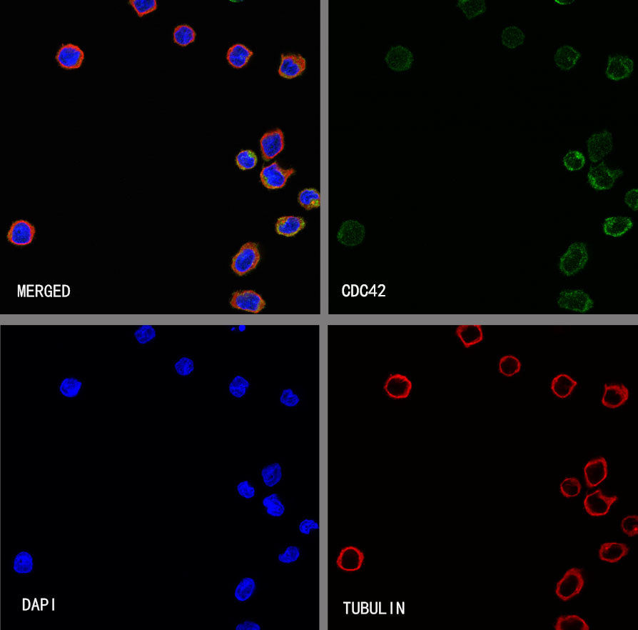
ICC shows positive staining in U937 cells. Anti- CDC42 antibody was used at 1/2000 dilution (Green) and incubated overnight at 4°C. Goat polyclonal Antibody to Rabbit IgG - H&L (Alexa Fluor® 488) was used as secondary antibody at 1/1000 dilution. The cells were fixed with 4% PFA and permeabilized with 0.1% PBS-Triton X-100. Nuclei were counterstained with DAPI (Blue). Counterstain with tubulin (Red).